Abdominal pain during pregnancy are very different, both in terms of subjective feeling as well as in terms of the location and intensity of occurrence. The pain can occur both at rest and after physical activity. The symptoms can occur in one place or spread to other areas.

- Leg exercises after standing work
- When should a doctor be consulted?
- Types of Leg Sprains
- causes
- Causes of hip dislocations
- The consequences of an injury.
- Treating a foot sprain – all doctors 84
- Which examinations are necessary?
- Where the doctor can refer you
- Related specialists
- Moscow traumatologists – opinions
- Frozen pregnancy
- Ectopic pregnancy
- TIPS FOR THE 26TH WEEK OF PREGNANCY
- Sixth month of pregnancy: changes in the woman's body and development of the fetus by weeks
- A healthy diet during pregnancy.
- Tests and examinations during pregnancy
- How to treat blood vessels on the legs?
- lifestyle
- symptoms
- diagnosis
- How the Fitbone intramedullary nail is implanted
- Exercises for the leg muscles
Leg exercises after standing work
If your legs hurt after work, this affects your usual quality of life. Exercise, pharmacological agents and traditional medicine can help solve the problem.
Different groups of factors can contribute to this symptom:
- A sedentary lifestyle: at work, in transport, at home. The load on the legs is reduced many times over and muscle tension is reduced. However, as soon as you have to walk longer than usual, your legs start to 'hum'.
- Standing work puts additional strain on the legs and causes pain and swelling in the ankles.
- Being overweight not only has a negative effect on the body, but also on the legs. The proportion of fat exceeds the proportion of muscle and the skeleton of the limbs cannot bear the load.
- Constantly wearing knee socks, stockings or tights. These compress the veins, which leads to blood congestion and nutrient deficiency. The solution in this case can be special healing stockings that restore blood circulation.
- Chronic diseases usually arise from lack of care, but if a person already suffers from varicose veins or joint and spine problems, legs will hurt more often.
- Vascular diseases: Blood vessels narrow and blood flow to muscle tissue is reduced, leading to phlebitis. The causes are varied, but occur more often in smokers, high blood pressure patients and people who have suffered a heart attack or stroke.
- Inflammatory processes in the joints and tissues of the legs, trauma.
When should a doctor be consulted?
Daily leg pain that is not caused by a sedentary lifestyle or, on the contrary, excessive stress at work requires treatment from the following specialists:
- Surgeons. A fracture, fracture or dislocation can be visible for a long time, so if an injury occurs, it is imperative to see a surgeon.
- Orthopedist. Corrects the various defects of the lower limbs and feet (flat feet, arthritis, arthrosis), which are a common cause of pain.
- Phlebologist. Deals with disorders of the vascular system.
To determine whether a doctor's visit is necessary, measure the diameter of your ankle in the morning before you go to bed and in the evening when you get home from work. If the difference in volume is more than 1 cm and the ankle is severely swollen, a visit to the doctor is mandatory.
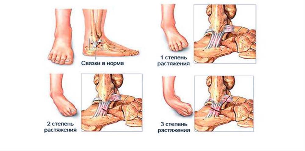
Types of Leg Sprains
- Easy (I). Characterized by a microscopic tear in the band. The injury is accompanied by mild pain and swelling of the injured joint. The person can move freely after 2-3 days without experiencing any discomfort.
- Moderate (II). Some of the fibers are torn. Mild pain and swelling occur.
- Severity (III). The ligaments are completely torn. This type of injury is characterized by acute pain and a sense of mobility, which is unusual for this joint. It requires long-term treatment followed by rehabilitation.
The ankle ligaments are more frequently injured. Only a traumatologist who conducts an examination using various diagnostic methods can determine the extent of the injury.
causes
An ankle sprain most commonly occurs when a person twists or trips while walking/running or as a result of a fall. This type of injury is common among athletes. The injury occurs when lifting weights, throwing a ball, or swinging your leg violently.
Other factors that promote a sprain:
- excessive weight;
- wearing uncomfortable platform shoes or high heels;
- Misalignment of the feet when jumping;
- diabetes;
- Age criteria – older people are at increased risk;
- Physiological features of the foot – flat feet, high ankle height;
- Falls on the ankle.
These injuries occur during strenuous physical activity due to strain on the lower limbs. Ligament injuries occur from fast jogging and sudden braking. The aforementioned causes are the most common. It should also be noted that dislocation can cause instability of the joint due to infectious lesions, pathological disorders and other previous injuries.
Causes of hip dislocations
To understand the causes of hip dislocations, one should remember to take a course in anatomy, physiology and kinesiology.
The hip joint is the union of two bones: the femoral head and the hip socket of the pelvic bone. The hip joint is surrounded by a variety of gluteal and thigh muscles that are connected to the bony structures via tendons and fascia. Thanks to this structure we are able to perform a wide range of amplitude movements in the joint: rotation, abduction, adduction, flexion and extension of the hip.
When we twist, bend or extend our limbs too much, the muscles and tendons are put under too much strain and can become injured - a major cause of overuse. In addition, the ligamentous system of the hip joint can be damaged:
The consequences of an injury.
Physical rehabilitation after a hip dislocation is a lengthy process. When it comes to hip ligament sprains, there is often no clear clinical picture. For many patients, a slight aching pain in the groin and limited mobility are not a reason to see a trauma surgeon. But not in this case!
Even the slightest stretching of the ligaments and muscles of the hip joint causes deformation of the cartilage and ultimately deformation of the bones. The bag This is followed by degenerative changes in the joint capsule. This already leads to deforming osteoarthritis of the hip, which in the advanced stage can only be helped by endoprosthetics. All of this can only happen due to a sprain that is not treated in a timely manner.
Treating a foot sprain – all doctors 84
Choose a date for online booking metro stations: Yugo-Zapadnaya Prospekt Vernadskogo
Which examinations are necessary?
Your doctor will tell you what tests you need to have done. You may receive an appointment for an examination:
Where the doctor can refer you
Services that may be recommended to you for treatment or further diagnosis:
Related specialists
To determine the most effective treatment, your doctor may refer you to a specialist:
Moscow traumatologists – opinions
I have an appointment with Dr. Roman Vadimovich agreed. During the consultation, the doctor listened to me very carefully, asked questions and showed me my pathology on the screen, which had an anatomical photo of the foot. He explained and showed me exercises. I enjoyed everything very much. Competent and professional approach to therapy. I recommend him!!!
I've never waited so long for an appointment for free!
Thank you very much!
Very good, attentive, friendly and sensitive specialist! I refused surgery after my injury and the doctor examined me, looked at all the scans and explained the consequences to me and together with the doctor we agreed that I would go to the hospital and have an operation.
He was an excellent doctor, he listened carefully to me and explained everything to me in detail. I was very pleased with the timing and the doctor created a treatment plan that no other doctor had done so well before.
My aunt was diagnosed with a sprain after riding her horse. She went to an orthopedic traumatologist and quickly got help. After the first treatment the pain had already subsided. The doctor did a good job.
A conversation took place. The doctor conducted a detailed consultation. He informed her about the treatment. He suggested various treatment options. He told me to do scans. I went and took pictures and showed them to him. He put me on the couch, touched my leg and made the diagnosis immediately, even without an ultrasound! Still, I went and had the scans done. He looked at it and confirmed there was fluid in the knee. Everything was explained and explained to me. The doctor performed the injections. I had the fluid and medication pumped out, a block, and he injected my knee. And when I arrived in Orekhovo-Zuyevo, my leg was already gone. I had been suffering since April. The doctor gave me recommendations for orthopedic shoes. Overall, I received very good feedback. I enjoyed everything very much. A detailed consultation. And immediately the opportunity to act. It is not necessary to take 100 tests and go to all the gynecologists and so on. No comparison with the doctors we have in Orekhovo-Zuevo. Especially in the polyclinic. I didn't like that there was no way to park the car. I had to go somewhere and park my car there. It was the first time we were there so it was an unexpected moment. We left the car and walked with a sore leg. The staff in the clinic communicated very well. I would definitely recommend the clinic and doctor to my friends. I came here on a recommendation. Two years ago a woman came here, also from Orekhovo-Zuyevo. Here in Orekhovo-Zuyevo they wanted to classify her as unfit for work. They wanted to give her the first group. Someone also recommended this doctor to her, Simonov AB. She came and he saved her hand. He's working on her. She says she should have come a year earlier. So we had a very good experience in Orekhovo-Zueva!
Frozen pregnancy
The fertilized egg does not always develop properly. In some cases, the egg cell stops dividing and dies. This is the most common case. An aborted pregnancy Through some kind of mutation. The woman is unaware that the pregnancy has been terminated.
However, the necrotic gestational sac begins to shed. This leads to long-lasting pain in the lower abdomen and soon afterwards to bloody discharge from the genital tract.
If a frozen pregnancy is diagnosed, treatment may be indicated. Conservative treatment is also possible, but can only be decided by a specialist after consultation.
Ectopic pregnancy
Most ectopic pregnancies are ectopic pregnancies in which the fertilized egg does not reach the uterus and implants in the fallopian tube. In this case, the development of the amniotic sac can continue for a long time without symptoms, even up to 12 weeks of pregnancy. However, in most cases the pregnancy is terminated after 6-8 weeks.
The fetus develops and grows, causing pain in the right or left side of the lower abdomen. The pain is one-sided, urgent and tends to get worse.
In addition to the abdominal pain, there is bloody discharge from the genital tract, and the pain radiates into the leg on the side on which it occurs. An unpleasant feeling of pressure may occur in the rectum. Medical intervention is the only way to preserve the woman's life. It is not possible to save the pregnancy.
TIPS FOR THE 26TH WEEK OF PREGNANCY
25-26 weeks of pregnancy is a good time to prepare for breastfeeding: wash your breasts with cool water, take air baths, gently massage your nipples with a stiff towel, but do not irritate them too much with it the uterus does not become tense.
A growing belly does not allow you to sleep. For greater comfort, take a small pillow to bed with you to place under your back, stomach and legs, or buy a special crescent-shaped pillow.
The 26th week of pregnancy is a time when rest is very important, but don't forget that exercise is also important for your health. If there are no contraindications, go for a walk, do gymnastics, but do not lift heavy weights. Now you need to be careful and watch out for falls, as they can trigger placental abruption, premature withdrawal of amniotic fluid and the onset of labor. Do not overexert yourself, and if you feel weak, lie down immediately. A squatting position can help with shortness of breath.
Watch your weight in the 25-26 weeks of pregnancy. Try to control your appetite and only eat fresh fruits and vegetables that are healthy for your baby.
Sixth month of pregnancy: changes in the woman's body and development of the fetus by weeks

Pregnancy and birth.
A healthy diet during pregnancy.
How to put together a healthy diet for pregnant women, which vitamins must be included in the diet, how much to drink, which drinks to prefer and which to avoid, which foods are harmful for pregnant women and how to keep your weight under control and at the same time stays in a good mood.
Tests and examinations during pregnancy

How to treat blood vessels on the legs?
The type of treatment for blood vessels on the legs depends on the diagnosis made by the doctor. You can find answers to the question of how to effectively treat unhealthy blood vessels on the legs from the doctors of the vascular centers in Moscow. There are two main areas in the treatment of vascular diseases of the legs:
- On the one hand, it is about the severity of the symptoms, such as: B. Pain in the legs, so that the patient can resume physical activity.
- Secondly, the progression of atherosclerosis is to be slowed down so that the risk of complications is reduced. Treatment without secondary prevention of atherosclerosis is not effective.
These goals can be achieved through lifestyle changes.
lifestyle
- Smoking cessation. This reduces the risk of complications of atherosclerosis of the lower limbs. Smoking causes artery damage and is considered one of the major risk factors for the development and progression of lower extremity artery disease.
If lifestyle changes are not enough, additional treatment is required. This is prescribed for serious diseases such as obliterative arteriosclerosis of the lower limbs. Your doctor will prescribe medications to prevent blood clots, lower blood pressure, lower cholesterol, and control pain.
symptoms
When you see your doctor, a patient with knee ligament inflammation may have the following symptoms:
- Acute pain in the knee. The pain first occurs with exertion, then at rest and becomes dull.
- In the case of kneecap ligamentitis, the pain is limited to the attachment point of the ligament, more often in the lower area.
- Damage to the deeper layers of the ligament as a result of inflammation only causes pain when there is deep pressure on the joint.
- Swelling of the area affected by the inflammation.
- Overheating of the joint.
- Restriction of freedom of movement.
As the inflammatory process progresses, the ligamentous tissue is gradually replaced by cartilage tissue, accompanied by degenerative-dystrophic changes. Ligamentosis develops, which is the logical continuation of ligamentitis. Some sources even equate the two diseases because the symptoms and complaints of a patient with both diseases are similar.
The differences in morphological changes consist in the inflammation of the tissue, which is reversible in ligamentitis, and in the irreversible pathological transformation of ligamentous tissue into cartilage in ligamentotosis.
Important: In ligamentitis, the altered ligaments are impregnated with calcium salts and 'ossified'.
In such cases, the joint's mobility is drastically limited - not only due to pain, but also due to morphological changes.
diagnosis
The final diagnosis is made based on the patient's complaints, the results of the physical examination and the instrumental examination methods:
- X-rays in two projections (show old fractures, ossification zones of the ligaments).
- Puncture of the joint.
- arthroscopy.
- MRI (shows thickening of the damaged ligaments).
- Ultrasound – particularly informative when examining the patellar ligament because it is close to the skin. Degeneration and deterioration of blood circulation – this can be determined by ultrasound of the kneecap.
MRI may show areas of ossification and calcification in the ligamentous tissue. This suggests that the inflammation of the ligament has already moved into the ligamentous phase.
We also recommend reading another article about osteophytes.
How the Fitbone intramedullary nail is implanted
Preparation for Fitbone® intramedullary nail rod implantation begins with a thorough examination that includes, but is not limited to, the following
- A detailed clinical examination
- Measurement of all lengths and axes of the lower limbs
- Comprehensive X-ray examination
- CT/MRI examination in a high-resolution CT scanner
The doctor and patient then discuss the desired treatment result together in detail. Based on all the information, the team of specialists determines the optimal bone separation zones and the most suitable Fitbone® intramedullary nail model. If necessary, an individual adjustment will be made tailored to the needs of the individual patient.
The specialists at the ZEM (Center for Corrective and Reconstructive Extremity Surgery) in Munich work according to the principle of 'reverse planning', meaning they start from the desired end result and then decide on the choice of surgical technique, the degree of distraction and the intensity of the physical Rehabilitation in the period after surgery. Once all recommendations are available, the patient has a few months to prepare for the operation and choose a date for the operation. Six weeks before admission, a second outpatient consultation takes place, during which all questions of interest can be clarified directly with the operating surgeon.
On the evening before the planned operation, the patient is admitted to the special partner clinic of the Munich Center for Corrective and Reconstructive Surgery ZEM - Hospital of the Brothers of Mercy in Munich. After initial laboratory tests and a discussion with the anesthetist, the individually adapted Fitbone® is implanted. The operation is performed under general anesthesia and can take up to three hours, depending on the difficulty. During the procedure, a special radiopaque mesh is used to ensure that the rod is perfectly aligned with the bone axis. After the operation, the patient is transferred back to the ward; a stay in the intensive care unit is usually not necessary.
Exercises for the leg muscles
The final part of the complex consists of exercises that are performed to fill the muscles with blood. The blood is intended to supply the muscles with the nutrients needed for recovery. These exercises further stretch the muscle fibers, allowing microscopic tears in the tissue to heal more quickly.
You can use a skipping rope, but you can also do without it. Stand straight with your feet together. Start with easy standing jumping jacks. Jump up and down for five minutes. Then stop and rest for 30 seconds. Attach weights (1-2 kg) to your ankles and jump for another 3 minutes;
Attach weights (2-3 kg) to your feet. Support yourself with your hands on the back of a chair or other stable object. Without bending your knees, raise your legs in different directions. Do not lower your feet to the floor. This way you increase the tension in your muscles and train them better. Do these moves for 5 minutes;
Extend your arms and stretch your neck. Place your hands on your waist. Stand on your tiptoes and walk across the room without bending your legs. Try to stretch your spine as much as possible while walking. Continue the exercise for 3-5 minutes.
Complement the exercise with water treatments - take a hot bath. Good nutrition is essential for rapid muscle recovery after physical activity. Include protein foods in your diet: fish, chicken fillet, lean veal, nuts, cottage cheese. Use fresh vegetable salads as a side dish. Vitamin complexes will not be superfluous.
Read more:- ligaments in the ankle.
- Rupture of the ligaments of the ankle.
- ligaments and tendons of the foot.
- Damaged ligaments of the ankle.
- Injury to the ligaments of the ankle.
- Sprained ligaments in the metatarsal area.
- Partial tear of the ligaments of the ankle.
- How long it takes for the ligaments to heal.