If you can, encourage your child to skateboard or snowboard. Clubfoot has no chance in these sports.

- clubfoot
- Clinical manifestations.
- Causes of clubfoot formation
- mechanical influences
- Nerve and muscle overload
- Toxic Effects
- genetic influence
- classification
- Clinical picture of congenital clubfoot in children
- Gymnastics, massage and footwear.
- By the way
- Treatment
- reviews
- Clubfoot diagnosis based on magnetic resonance imaging and computed tomography of the feet
- methods of choice
- What an x-ray examination of the clubfoot shows
- Typical manifestations or signs of clubfoot in children
- 3 forms of congenital hip dysplasia
- What are the symptoms that indicate a child might have hip dysplasia? How can it be diagnosed?
- Symptoms of flat feet in children
- Treatment of flat feet in children
- diagnosis
- Modern methods
- massage
- An appeal to parents
clubfoot
This congenital or acquired ankle deformity is called clubfoot. Characteristic of this condition is the inability to fully plant the sole on the ground, and the deviation looks like the foot is turned inside out. The developmental anomaly of the musculoskeletal system, or clubfoot, which takes a long time to treat, can appear at any age.
The true causes of childhood clubfoot are not known, but there are theories about the occurrence of the condition with certain predisposing factors:
- Malposition of the fetus during pregnancy – the problem is related to the pressure of the uterus on the developing skeleton, which causes an imbalance in the blood supply to the baby's lower limbs;
- the use of certain medications by the pregnant woman;
- insufficient development of the musculoskeletal system;
- hereditary predisposition – a problem in one parent increases the risk of developing clubfoot.
Abnormalities in adolescence and adulthood include.
- insufficient bone healing after a bone fracture;
- burns in the lower leg and foot area;
- various bone diseases;
- neurological disorders;
- malignant and benign tumors.
Abnormal bone growth plays a major role.
Clinical manifestations.
The main symptoms of clubfoot include:
- Lowering the outer edge of the foot while elevating the inner foot;
- position of the feet at an angle to each other;
- leaning on the outside of the foot;
- outward displacement of lower leg bones;
- Rotation of the foot with the sole upwards.
Secondary symptoms of clubfoot before treatment are shown:
- Minimum foot size;
- Complete absence or marked limitation of ankle mobility;
- transverse flexion of the foot;
- Raised heel with toes pointing down;
- Inward displacement of the heel.
In acquired clubfoot, the symptoms in children aged 2-3 years before treatment are:
- Changes in gait pattern without recognizable cause;
- Abnormal foot position - the foot is visible in the sand or snow and the feet face each other;
- Abnormal alignment of the knee joints;
- Mobility problems in the feet - obvious limitation of movement;
- Inclination of the big toes.
Clear signs of the disease can be seen with complete relaxation or during sleep.
Causes of clubfoot formation
There are the following medical factors that predispose to clubfoot and are involved in the development of congenital clubfoot disease
mechanical influences
Caused by a misalignment of the baby in the womb. The wall of the uterus presses on the fetal ankle, causing poor blood flow and deforming the baby's limbs.
Nerve and muscle overload
Causes: Abnormal development of ligaments and muscles that affect the development of the child's foot.
Toxic Effects
Causes: Early drug treatment by the mother.
genetic influence
According to medical opinion, a congenital pathological process is due to a genetic predisposition to its occurrence. However, this is not always the case as the disease can also be acquired. In the latter case, the causes of the disease are different: with abnormal healing of ankle fractures, deep burns in the ankle area, neurological diseases, tumors and pathologies related to the child's growth retardation.
classification
The classification of the different types of clubfoot is based on the etiology and severity of the condition. Depending on the origin of the disease, a distinction can be made between the following forms.
- Idiopathic clubfoot that is primary;
- Secondary clubfoot as a result of unfavorable fetal factors during prenatal development.
REFERENCE! Secondary clubfoot is a consequence of pathologies such as spinal hernia, arthrogryposis, myelodysplasia, amniotic band syndrome or immobile dura syndrome.
Depending on the severity of the disease, the classification divides congenital clubfoot into mild pathology with the possibility of full correction, moderate clubfoot and severe deformity, in which the lesion is particularly pronounced and characterized by high rigidity.
By far the best fitted classification is that proposed by Ponseti. It is of practical value in distinguishing and treating the disease. The author distinguishes the following types of pathologies:
- clubfoot uncorrected before age 8;
- corrected Ponseti clubfoot;
- Recurrent pathology;
- clubfoot unresponsive to treatment;
- Atypical clubfoot.
The treatment of these types of clubfoot is fundamentally different. Ponseti's classification, while one of the best, is not an exact classification.
Clinical picture of congenital clubfoot in children
According to current scientific opinion, congenital clubfoot is based on profound structural changes in the tendons, muscles, capsule and ligaments of the foot. Therefore, the severity of clubfoot deformity depends on the extent of changes in these tissues and the severity of the deformity.
In order to correctly assess the severity of the foot deformity in a typical congenital clubfoot, three grades can be distinguished:
Mild degree Congenital clubfoot is manifested by the presence of all the elements of the deformity, which can be easily corrected with manual corrections.
medium grade – has all the components of a congenital clubfoot, but with manual correction some elements of the deformity remain and are difficult to preserve with cast or other fixation devices.
severity – All components of congenital clubfoot are severe, cannot be corrected by manual techniques, and are divided into soft-tissue and bony forms according to age and severity of manifestation.
The clinical picture of the typical form of congenital clubfoot consists of three main elements:
- lowering of the foot or plantar flexion (equinus);
- Anterior and medial inward flexion of the foot (adduction);
- Rotation of the foot about its longitudinal axis with the inside of the foot pointing up and the outside of the foot pointing down (supination) (photo).
The mentioned elements of the clubfoot increase with the age of the child and there are secondary changes in the mentioned segments; Especially in unilateral clubfoot, shortening, tibial rotation with valgus deformity at the knee joint, pelvic obliquity and other static anomalies develop in addition to the above-mentioned elements. The clinical picture of bilateral clubfoot is characterized by the greatest functional impairment. Children with bilateral clubfoot cannot stand in one place and are constantly moving in small steps, bending their knees and leaning their bodies forward for balance. Children with severe clubfoot walk strenuously with their feet spread, placing one foot on top of the other, primarily stressing the lateral pelvis of the foot, and this posture quickly leads to fatigue.
Gymnastics, massage and footwear.
The internal alignment of the feet when walking is an easy problem to solve and does not require special treatment, but work needs to be done to put the child's feet in the right position.
If clubfoot is caused by increased muscle tone, special exercises should be performed. A podiatrist can tell you what exercises these are.
Perform a massage regimen to relieve the increased tension. The massages are prescribed by a neurologist.
Bathe your child in a salt bath every other day. It strengthens the whole body and relieves hypertension. Dilute 3-4 tablespoons of liquid pine extract and a glass of sea salt (unscented, available only at pharmacies) in a bucket of water. If the child is less than a year old, take half a glass of salt. Bring the water to a temperature at which the child is used to bathing. After the salt-iodine bath, the child should be bathed in fresh water.
By the way
Orthopaedists are against it! Parents are happy when a baby tries to walk at the age of 9-10 months. This is how a little rascal grows up! Some people even encourage babies to walk early: they hold their hands, buy a walker. Orthopedists categorically reject this. Both the muscles and the skeleton should be large enough for the child to walk. All city kids today have a mild form of rickets. When a city kid starts walking too early, they're bound to have crooked legs and crooked feet—their bones can't take it. So there's no reason to rush anything. If your child still has difficulty walking at 10 months, you should give him an extra massage to strengthen his muscles.
bad advice. Advice mothers give each other for switching clubfoot babies' places: Put right shoe on left foot and left shoe on right foot - bad. The consequence is that the child not only does not have straight feet, but also twists his legs in the knee and hip joints.

Squinting in children is one of the most common malpositions of the musculoskeletal system. If left untreated, it can have devastating consequences, such as: B. Inability to walk and lifelong disability.
– Over the past five years, the number of young patients diagnosed with clubfoot has roughly doubled in our region. We have to take care of three to four babies a month,' says orthopedist and traumatologist Dmitry Ibragimov. – A few years ago, in one of the clinics, I saw a one-and-a-half-year-old boy with his legs on his side. I invited his mother to visit: 'Everything is fine', the woman said. – My father walked like that when he was a child. '. I persuaded her to have an exam and the boy showed the first signs of clubfoot. The child was treated successfully, but he could have overlooked it….
The causes of clubfoot are still poorly understood. Foot curvature can be caused by compression of the foot during fetal development. Abnormal and delayed formation of foot and lower leg. Deformation of the nervous system.
Treatment
Treatment and whether it can be done at home is decided by the podiatrist after diagnosis. The earlier therapy is started, the better the chances of eliminating the anomaly. Parents should be aware that Clubfoot cannot be treated quickly and the therapy requires a consistent and careful approach in close cooperation with the attending physician.
Many parents wonder if clubfoot will have consequences. This depends on the extent of the anomaly. If the degree of congenital clubfoot is small, the treatment is very effective in 95 % of cases: full correction of foot position at home is possible without surgery. In severe clubfoot, conservative treatment is statistically effective in only 10% of cases.
In the case of congenital clubfoot, it is advisable to start treatment in the first few weeks of the child's life. At this point, the bones are still soft and flexible, and the child can be treated without surgery. Correct alignment of the foot can be achieved through gymnastics and massage. For infants, gentle immobilization of the feet in the correct position - bandaging and taping - is often recommended. If the pathology is severe, a graded cast is applied.
At a later stage, children are always prescribed im. Physiotherapy, massage, physical therapy and orthopedic shoes are recommended.The child should wear orthopedic shoes specially made for the child, taking into account the recommendations of the orthopedist.
If conservative treatment does not work or the results do not satisfy the parents and the doctor, surgical correction of the child's foot may be recommended at the age of 1-2 years. This is usually plastic surgery on the tendons and ligaments. After the surgery, the child must wear a plaster cast, which can last up to 6 months or more.
Tendon and ligament surgeries are usually performed on children from 1.5 to 2-3 years of age, as well as children from 4, 5 and 6 years of age. From the age of 7, operations on the bony foot apparatus or, if necessary, combined operations can be performed.
reviews
There are not only many testimonials from parents about clubfoot treatment on the Internet, but also entire social media groups where mothers share their experiences with other parents. Treatment of a foot anomaly in a child is long, difficult and in each case requires a very individual approach, so someone else's experience is not always helpful. Even massage and gymnastics can be different in different children, since in one child the anomaly is caused by pathology of the ligamentous apparatus, and in another - neurogenicity.
The treatment usually lasts from several months to several years. And during this time, mothers must maintain faith in the success of the treatment. This is sometimes difficult, and sometimes despair sets in. This is actively exploited by shady people who go to such forums and parent groups and actively advertise some 'specialists' who, with their hands and herbs alone, can fix children's feet in a few weeks without plaster or surgery. Does not work like that. And these 'specialists' are usually only concerned with the parents' wallets.
Watch the following video about congenital clubfoot.
Elena Lisova
Medical assessor, specialist in psychosomatics, mother of 4 children.
Clubfoot diagnosis based on magnetic resonance imaging and computed tomography of the feet
Classification: Congenital clubfoot, clubfoot formation, congenital equinovarus clubfoot, neurogenic clubfoot, and clubfoot arthrogrypsis.
methods of choice
- Radiological examination in two projections (dorsal and lateral)
- The radiological examination is not carried out at birth, but only after 4 months. After splinting to determine a possible indication for surgical treatment or to plan a surgical intervention
- To obtain an image of the foot in maximum dorsiflexion (assessing Achilles tendon shortening).
What an x-ray examination of the clubfoot shows
- Image of the foot in the corrected position
- Dorsal projection is less than 15° (typical 15-40°) and lateral projection is less than 25° (typical 25-45°)
- Subluxation of the navicular bone (dorsal view)
- Ossification nuclei in the talus, heel, elbow, and metatarsal bones are always visible at birth
- Ossification of the nucleus pulposus is not visible in the navicular bone (ossified at 3 years of age), requiring careful assessment of navicular subluxation.
- The position of the ankle can be assessed by determining the angle between the axis of the talus and the first metatarsal.
- Tibial carpal angle greater than 90° (normal 60-90°)
- Back-to-front inclination of the heel bone
- Adduction Deformity: In the distal view, the talar axis is lateral to the base of the first metatarsal.
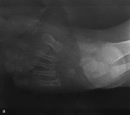
a, b crookedness at 5 months of age: a) dorsal radiograph shows shortening of the Achilles tendon, lack of posterior-to-anterior tilting of the calcaneus, and horizontal involvement of the talus resulting in a reduced angle of the talcaneus;
Typical manifestations or signs of clubfoot in children
- A palpable anterior edge of the heel bone
- Thin skin with fine wrinkles in this area
- Shortened Achilles tendon, palpable like a tight Achilles tendon
- Lateral ankle shifted backwards
- Low heel height
- Thin calves
Four stages of treating clubfoot in children:
– Level I: chiropractic manipulation and cast splints (immediately after birth).
– Phase II: surgical removal of the periarticular contracture (at the age of 4-6 months).
– Phase III: Splinting (if the foot remains in adduction position).
– Phase IV: Correction of recurrent deformities and late bone displacement problems.
Treatment of Clubfoot with Manual Manipulation and Cast Splints (Stage I)
An uncomplicated clubfoot can be adequately treated with treatment levels I and II.
An important part of all stages of treating children's clubfoot is a comprehensive massage and the right choice of shoes. The choice of corrective shoes for clubfoot is made in consultation with a podiatrist, after an examination and appropriate podometric and plantographic tests.
3 forms of congenital hip dysplasia
- articular dysplasia, That is, it is due to the immaturity of the joint. The structure of the acetabulum is disrupted, causing the femoral head to shift.
- epiphyseal plate. It is a joint stiffening that leads to a deformation of the limbs. The whole thing is accompanied by a pronounced pain syndrome. During an ultrasound examination of the child, these disorders are clearly visible.
- Rotation. An anatomical misalignment of the bones that causes the child to have a club foot.
Also, the dysplasia can be mild or severe. For this reason, it is further subdivided into grades:
- Grade 1 is premature dislocation. This is the most common grade. Over time, the pathology may spontaneously regress or progress to the next grade.
- Grade 2 - Subluxation. The ligaments are stretched and lose tension.
- Grade 3 - Dislocation. The femoral head is displaced and completely exits the acetabulum.
What are the symptoms that indicate a child might have hip dysplasia? How can it be diagnosed?
A neonatologist or pediatrician will examine the child in the hospital. If there are visible changes, they may recommend additional testing to make a diagnosis. Nobody can rule out that there are not immediately visible signs of anomalies, but they only appear months later.
Therefore, it is important to visit the specialist several times a year so that you do not overlook the following symptoms:

Asymmetry of the buttocks, groin and thigh folds
How to recognize it. Lay your child on their back or stomach and stretch their legs slightly. Notice how the skin folds are in the groin, on the thighs and under the bottom. Most importantly, the pleats are the same size and at the same angle.
your baby's knees are not the same size
To find out, lay your child on their back, straighten their legs, and then bend their knees (they should be even). Is one knee higher or lower than the other? It is likely that your child's joints are at different heights.
The joints have different amplitudes when you move your legs apart.
You can determine this by laying your child on his back, bending his legs at the knees and spreading them apart. Children up to 1 year have good mobility, even without strength they can stretch their hips so far that they simply 'lie down'. Your baby definitely has hip dysplasia if you hear a pop or the amplitude of hip movement is different.
IMPORTANT!!! If your doctor or you diagnosed your baby with dysplasia before the age of 6 months, there is a good chance of a full recovery.
There are a number of other ways that can help identify hip dysplasia in your baby. This includes:
Symptoms of flat feet in children
Parents often do not know that their child has flat feet. In most cases, it does not bother the child, but sometimes he complains of pain and discomfort when walking.
The following symptoms may indicate foot abnormalities:
- Quick fatigue – the child prefers to sit on a bench than actively participate in games with peers, quickly gets tired when running or on long walks.
- Pain in your feet (sometimes there may be pain in your knees or lower back).
- lameness after long walking.
- Uneven wear of the footwear (the sole only wears out on the outside or inside).

Treatment of flat feet in children
In order to detect foot problems early, children should visit a podiatrist regularly, ie once a year, for an examination. This is especially important if the child complains of pain and discomfort when walking.
diagnosis
The diagnosis of flat feet is made during a visit to the orthopedist: the doctor carefully examines the child, establishes a family history of flat feet and, if necessary, prescribes a plantography (in the past, the child's feet were smeared with dye, an impression was made on paper, and then examined, today the recording and processing is done with the help of computer technology). The official diagnosis of flat feet is made only after an X-ray examination of the foot, which reveals not only the presence, but also the type and degree of flat feet in children.
– Before the age of 7, a flat foot is generally not diagnosed. The child is growing steadily and the feet are also developing. Many children have an axillary foot deformity that also affects the foot. Usually, around the age of 5 or 6, this pelvic tilt goes away and the foot straightens out. So when I hear that a child is diagnosed with flat feet at the age of one, I think that's wrong,' says orthopedic surgeon Roman Żaryn.
Modern methods
– When treating flat feet, the most important question is whether it is the flat feet that are bothering the child or if it is a problem that is only bothering the child's parents. The main treatment is aimed at prevention: therapeutic massage, physical therapy, physiotherapy (electrostimulation and paraffin compresses), but one should not expect global improvement from physiotherapy, says the doctor.
massage in the treatment of children with flat feet is effective because it restores blood circulation in the foot and has a beneficial effect on the joints and ligaments. It is best to leave the massage to a specialist, especially if the child has suffered a serious sprain or fracture. A massage mat can be used at home as it works on pressure points in the foot and can stimulate blood circulation.
massage
Before you start exercising at home or in the gym, it makes sense to loosen up the problem muscles that you've already become familiar with. Buying literature on massage can help you become familiar with the techniques and proper execution. I was taught by VI Vasichkin 15 years ago. I massage children's backs and feet, concentrating on relaxing problem muscles. You can also use the books 'Children's Massage' and 'Flat Feet' by Irina Krasikova, in which you will find not only massage techniques, but also a set of exercises.
It is good if you have the opportunity to work out in a gym, but it is not a requirement. You can train outdoors, in the forest, in nature and of course at home. You should work out the exercises, systematize them, gradually increasing the load. You can talk to the physical education teacher at school who can suggest some exercises for your child.
Once you understand what needs to be done and explain it to the child, start doing the exercises together with him. For the second hour, bring a video camera and film the child's awkward movements.
After 5-7 sessions you will see the first results. Show your child the video of the lesson. The children are usually happy about their progress, are proud of themselves and absolutely want to get ahead. Don't be fooled though, you are just at the beginning of your journey. Don't forget to praise and encourage your child.
So you've started. It is best if your child concentrates as much as possible on what he is doing. Run a few laps first, this warms up the muscles and most importantly helps you to recognize the problem again, especially in the corners. From that day on, your child will no longer walk as he feels comfortable. It will now learn to run and walk in a new way.
When running, it's not about speed, it's about the right foot position. In the next session, introduce a system of 'extra exercises' for inattention and foot deformities (remember the claps and push-ups). For the first few weeks, you need to pay close attention to what your child is doing, rubbing their hands together to clap.
An appeal to parents
My solution is: You noticed the problem, you saw a doctor, a trainer, a massage therapist, you started forming new habits, you relaxed tight muscles and engaged antagonistic muscles, you made your child believe that it's great. You have now opened the door to sport and a healthy life for your child.
May your children always be healthy, beautiful and strong!
The information on this page is for informational purposes only and is not a recommendation for self-diagnosis and treatment. Always consult your doctor if you have any medical questions.
Read more:- What is clubfoot?.
- Congenital clubfoot.
- The baby has short legs.
- Photo of scraped feet.
- Why does a child develop clubfoot?.
- Baby with clubfoot in shoes.
- Clubfoot in 7-year-old children.
- clubfoot.