Print this article

- Arthrodesis of the tarsal joint
- Ask a question
- When will doctors stop using this technique?
- Indications for arthrodesis
- keywords
- Types of arthrodesis
- Painkillers during arthrodesis
- Indications and contraindications for knee arthrodesis
- Implementation technique.
- Arthrodesis options
- Indications and contraindications for arthrodesis
- Arthrodesis of the ankle joint
- Osteoarthritis of the pelvic-thigh joint
- Three-joint arthrodesis
- Compression arthrodesis
- Surgical technique
- Rehabilitation after arthrodesis
Arthrodesis of the tarsal joint
It is now possible to activate an additional profile for a child in the client's personal practice. This can be done in the 'Settings' section of the personal practice if the child has already been treated at MEDSI.
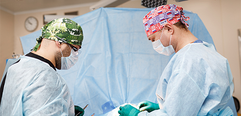
The orthopedic traumatologists in the inpatient department are specialists in this field. They deal exclusively with foot surgery, knee arthroscopy (anterior cruciate ligament repair and meniscectomy) and knee arthroplasty. This ensures the highest possible quality of these interventions. The department's doctors are trained at the best clinics in Russia and abroad and themselves teach doctors in Russia and neighboring countries.
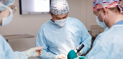
Doctors use Arthrex power tools to perform surgery on small and large joints. They are thoroughly tested in the company's quality assurance department and are characterized by the highest hardness and corrosion resistance. The procedures are carried out in a well-equipped operating room with a special operating table for trauma orthopedics and an arthroscopic frame from Karl Storz. Arthrex and FootDoctor implants are used for foot and ankle surgery, and Arthrex implants are used for knee surgery. All implants are characterized by optimal performance characteristics (easy insertion, guaranteed biocompatibility, long service life). All these features ensure a high level of performance, safety and effectiveness even in the most difficult cases.
Techniques such as percutaneous and open foot surgery are used for operations on the foot and ankle: arthroscopy and arthrodesis. Knee reconstruction includes arthroscopic anterior and posterior cruciate ligament plasty, collateral ligament plasty, arthroscopic meniscectomy and patellar stabilization for dislocated knee joints. Diagnostic arthroscopies, unicompartmental and total knee arthroplasties are also performed. The techniques used guarantee a quick procedure and a short recovery time after the operation. Rehabilitation of patients is possible in the MEDSI sanatorium in Otradna. This modern rehabilitation and wellness center, just six kilometers from Moscow, offers everything you need for good rehabilitation and relaxation. It has excellent therapeutic and diagnostic facilities and uses its own medical methods.
Ask a question

This procedure replaces the components of a dysfunctional joint with an endoprosthesis and allows:
Knee joint replacement is performed when:
- Osteoarthritis in stages 3 and 4
- Osteoarthritis of the tibia and femoral condyles
- Cystic condylar transformation
- rheumatoid arthritis
- Tumor processes
The operation takes about an hour. The patient stays in the hospital for 4 days, the stitches are removed on day 14-16. 1.5-2 months after the operation, the patient can walk without additional support.
Foot surgery is often performed for problems such as hallux valgus (bunion, a 'lump' on the foot). This problem involves an outward deviation of the big toe. When it develops, the patient not only experiences pain when standing or walking for long periods of time, but also difficulty adjusting shoes. The bone can only be removed surgically. Thanks to modern techniques, you can get back on your feet immediately after the correction. Our surgeons eliminate the deformity and restore the function of the joint.
The following techniques are used in foot surgery:
- Percutaneous surgery. This surgery is minimally invasive and allows bone to be removed through small incisions.
- Reconstruction of the forefoot. This procedure is performed when the deformity has progressed. It is an open operation in which the soft tissues are incised, the axis of the toe is exposed, and the growths are removed.
On average, each procedure only takes 30-40 minutes. A plaster cast is not required after the procedure. At the same time, the operation restores the aesthetic appearance and full functionality of the foot.
When will doctors stop using this technique?
If the treatment of the joint with this technique proceeds normally and without complications, stability of the knee joint and the possibility of full weight bearing can be achieved, allowing patients to have an overall satisfactory quality of life. Recent studies also suggest that this method can combat mixed infections or infections caused by highly virulent microorganisms when other surgical techniques have failed.
Arthrodesis has proven effective in patients with multiple risk factors for reinfection of the prosthesis and at the same time lower functional demands on the knee joint. It is the only effective treatment method if it is possible to at least keep the joint as a body support.
Indications for arthrodesis
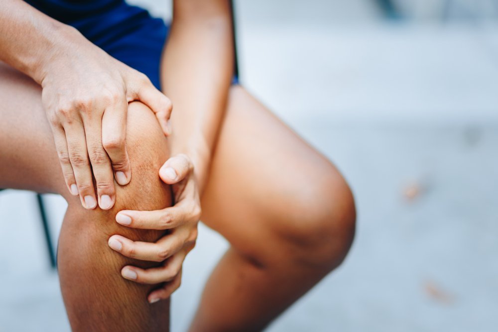
It is indicated for all joint diseases in which the joint surface is damaged and every movement is painful.
- Severe deforming arthritis
- Bone lesions caused by osteoarthritis.
- Changes in the joint caused by poliomyelitis
- Severe contractures of the joint
- Hypermobility of the joint and its laxity due to atrophy of the ligamentous muscular apparatus.
- The patient is unable to move
- Joint involvement as a result of tuberculosis
- Wrong joint as a result of a fracture
keywords
tibia; fibula; autograft; ankle joint; post-traumatic arthrodesis; arthrodesis; intramedullary fixation
Yezhov MYu. Foot. Degenerative and dystrophic diseases of the joints of the foot and ankle. Nizhny Novgorod, 2011. p. 12-13. Russian (Ezhov MY Foot. Osteoarthritis of the foot and ankle. N. Novgorod, 2011. С. 12-13)
Omelchenko TN. Ankle fractures and rapidly progressive osteoarthritis of the ankle: prevention and treatment. Orthopedics, traumatology and prosthetics. 2013; (4): 35-40. Russian (Omelchenko TN Ankle fractures and rapidly progressive osteoarthritis of the ankle: prevention and treatment // Orthopedics, Traumatology and Prosthetics. 2013. № 4. С. 35-40)
Saltzman CL, Salamon ML, Blanchard GM, Huff T, Hayes A, Buckwalter JA, et al. Epidemiology of ankle arthritis: report of a consecutive series of 639 patients from a tertiary orthopedic center. Iowa Orthop J. 2005; (25): 44-46
Gorbatov RO, Gorin VV, Pavlov DV, Malyshev EE. Competence of modern ankle arthrodesis for post-traumatic crusarthrosis, grades III-IV. Modern Technologies in Medicine. 2016; 8(3): 64-74. Russian (Gorbatov RO, Gorin VV, Pavlov DV, Malyshev EE Concept of modern ankle arthrodesis for post-traumatic crusarthrosis grade III-IV // Modern Technologies in Medicine. 2016. Т. 8, № 3. С. 64-74.) DOI: 10.17691 /stm2016.8.3.07.
Grazhdanov KA, Barabash YuA, Norkin IA, Zuev PP, Kauts OA, et al. Results of surgical treatment in patients with sequelae of metaphysis injuries of the distal tibia. In: Proceedings of the Ilizarovskiye Chteniya conference. Kurgan, 2021. pp. 86-87. Russian (Grazhdanov KA, Barabash YA, Norkin IA, Zuev PP, Kauts OA, et al. Results of surgical treatment of patients with sequelae of injury to the distal tibial metaphysis // Materials of the International Scientific and Practical Conference 'Ilizarov Readings'. Kurgan, 2021. С. 86-87)
Types of arthrodesis
The surgical procedure is divided into several types depending on which technique is used to immobilize the affected joint. The basic idea in all of them is to immobilize the joint. This can help stabilize the joint and reduce pain intensity.
- Intraarticular. In this case the joint capsule is opened. The surgeon then cuts out the superficial bone elements and removes the damaged hyaline cartilage. The bones are then placed in the most comfortable position and fixed with special metal pins or screws.
- Extra-articular. In this case it is not necessary to cut out the cartilage. The bones of the joint are fixed with a special bone graft.
- Combination. This technique combines all the techniques that the surgeon performs during intra-articular and extra-articular arthrodesis. First, the joint capsule is opened and the superficial bone elements are resected. The non-viable hyaline cartilage is then removed and replaced with a special autograft. The joint is immobilized using metal plates.
- Compression. The special thing about this method is that no transplants are used to immobilize the joint. The articular surfaces are compressed using a compression or compression-distraction apparatus (Grishin, Ilizarov, Volkov-Oganesian).
Painkillers during arthrodesis
Arthrodesis is never performed under local anesthesia as this is not enough to completely anesthetize the limb. Since the procedure is carried out on bony and cartilaginous structures, more global methods are used. The most commonly used methods are endotracheal anesthesia or spinal anesthesia. In the first case, the patient is put into a medically induced sleep in which he feels and sees nothing. In the second case, the lower limbs are numb, but the patient is awake and can see everything.
In some cases, combined anesthesia is used, which combines spinal anesthesia with medically induced sleep. This option is usually used for very anxious patients who are terrified of the operation.
Ankle arthrodesis takes between 2 and 6 hours. The duration depends on the size of the procedure, the initial state of the anatomical elements that make up the joint and the need for a transplant.
Indications and contraindications for knee arthrodesis
The main indications for such surgical intervention are various types of joint pathologies, which are accompanied by destructive changes in tissues and a pronounced pain syndrome.
Knee arthrodesis may be indicated for severe deforming arthritis with limited limb function. It is also indicated for certain types of osteoarthritis, severe joint instability and spastic flexion contractures. This treatment method can be used if a previous tuberculous infection has caused significant joint lesions.
Contraindications include:
- Acute inflammatory processes in the joint area;
- Purulent inflammation of the skin and soft tissues in the area to be treated;
- Systemic diseases in the decompensated stage;
- general acute infections, etc.
Implementation technique.
There are currently several types of arthrodesis. First, a distinction is made between extra-articular and intra-articular procedures. Procedures that affect the extra-articular joint are called joint fixation using a periarticular prosthesis. During intra-articular arthrodesis, the joint surfaces are removed. In some cases these techniques are also combined.
During arthrodesis, the joint is often fixed using a device such as the Ilizarov apparatus.
The arthrodesis technique is developing rapidly. It is now possible to carry out the operation using computer navigation. In 2013, scientists from Russia's Ilizarov Scientific Center for Reconstructive Traumatology and Orthopedics published the results of the operation. In 2013, scientists from Russia's Ilizarov Scientific Center for Reconstructive Traumatology and Orthopedics published a paper proving the effectiveness of the technique.
Before the operation, the patient undergoes general or spinal anesthesia. After immobilizing the joint, the wound is sutured and a drain is placed. A plaster cast is then applied for up to five months.
Arthrodesis options
Today we can define four main options for arthrodesis:
This operation involves opening the joint capsule and removing the hyaline cartilage and sometimes the synovial membrane. The bone is immobilized using various metal structures, such as: B. special plates. This variant of the operation is the most commonly used.
The joint capsule is not opened and the cartilage is not removed. The joint is fixed with a special bone graft. This procedure is most often used for joint damage caused by tuberculosis.
In this case, both intra-articular and extra-articular techniques are used. The cartilage is removed and a bone graft or metal plate is inserted.
To immobilize the joint, special compression and distraction splints, such as B. the Ilizarov rail is used.
Another technique is minimally invasive arthroscopic arthrodesis. In 2013, researchers at Acad. NN Burdenko concluded that arthroscopic arthrodesis of the ankle has a number of significant advantages over open surgery.
Indications and contraindications for arthrodesis
The main indications for arthrodesis are:
- Osteoarthritis in stage 3-4, when endoprosthesis is not possible;
- Severe deforming osteoarthritis;
- Persistent pain in the joint, even with light exertion;
- Significant functional impairment of the joint;
- Irreparably healed fracture or long-term dislocation;
- Severe flexion and extension contractures of the joint and many others.
Contraindications include age under 12 years, fistulas of unclear origin, purulent arthritis, hypersensitivity to anesthetics, decompensated chronic diseases, tendency to thrombosis, etc.
Arthrodesis of the ankle joint
The procedure is carried out via an open approach to the joint under general or spinal anesthesia and takes 2 to 3 hours. The surgeon opens the joint and removes cartilage and osteophytes (painful bone growths). In difficult cases, the joint is completely removed and the bones are fused at an appropriate angle (this can result in a shortened limb). Incisions are made on the bone surfaces, the bones are compressed at an appropriate angle and locked. The bones can be held together using screws, spokes, special metal plates, or bone implants. Most often, an intra-articular fusion is performed, only in some cases (in tuberculosis, when the joint capsule cannot be opened) an extra-articular one. If an arthrodesis of the ankle joint was performed, rehabilitation lasts 6-8 weeks. It requires physical therapy, massage and orthopedic resection.
It is indicated for severe fractures or dislocations of these bones, osteoarthritis, clubfoot, valgus foot and clubfoot. The goal of the operation is to fuse the talus and heel bones. During metatarsophalangeal joint arthrodesis, the phalanx of the toes is fused to the metatarsal bone. It is usually performed on the first toe (when a bone lump grows). Sometimes both phalanges are fused, with the toe completely immobilized in a slightly flexed position.
Osteoarthritis of the pelvic-thigh joint
It is caused by flat feet or deterioration of the ligaments due to excessive stress (obesity, standing work, high-heeled shoes). Over time, deforming osteoarthritis of the ankle develops, after which a fusion operation is unavoidable. Severe cases of flat foot deformity (when the inner part of the heel is worn down) require triquetral arthrodesis to form the proper arch of the foot. In severe cases, panarthrodesis (total foot arthrodesis) is required, that is, a simultaneous arthrodesis of the ankle, talus, scaphoid and calcaneus, in which the foot is completely immobilized.
Treatment and rehabilitation after joint surgery must be closely monitored by an orthopedic surgeon!
Three-joint arthrodesis
Anatomically speaking, a three-joint arthrodesis cannot be considered an ankle fixation because it immobilizes all three joints of the foot: the scaphoid, navicular, and talar joints. However, in many traumatology textbooks this procedure is described together with other ankle arthrodeses due to similar indications and end results. The procedure is most often performed for congenital and paralytic deformities, provides good stabilization of the foot and can be combined with other techniques (e.g. tenodesis).
The procedure is carried out under general anesthesia in the supine position. The incision begins behind the lateral ankle joint and ends at the level of the second sphenoid bone. The underlying tissue is cut, the fascia is severed, the joint ends are resected and the articular cartilage is completely removed. Check the correct alignment of the foot after resection of the bony structures. Make incisions on the bony surfaces that touch. Fill the empty space with small grafts from the resected areas. The wound is stitched up and a plaster cast is applied for 8 weeks.
Compression arthrodesis
This type of arthrodesis is performed using a compression or compression-distraction device (Grishin device, Ilizarov device, etc.). This technique allows for a reduction in the surgical volume, provides reliable stabilization, and shortens the time required for bone callus formation. The operation is carried out under general anesthesia in the supine position. The ankle joint is opened forward, the foot is dislocated, the joint surfaces are resected, cartilage is removed, and incisions are made at the mating ends of the bones. The spokes are inserted through the shinbone and foot bones and the splint is adjusted. In the postoperative period, additional compression is applied (1 mm for 7-10 days). Walking begins on the second day, full support is allowed from the second week, and the splint is removed after two months after ankylosis is radiologically confirmed.
The operation is performed through small incisions using special arthroscopic equipment. This method reduces trauma, rehabilitation time and the number of complications in the postoperative period. The anesthesia is carried out under general anesthesia or by the attending anesthesiologist. The joint is accessed through several incisions. The most common approach is the anteromedial incision. Other approaches depend on the technique chosen and the type of lesions. Tractions and special splints are used during the procedure.
Arthroscopic arthrodesis consists of three steps. First, the hyaline cartilage and adjacent bone fragments are removed. The foot is then brought into a neutral position and fixed with two or three screws. One screw connects the talus to the tibial metaphysis and the other to the fibular metaphysis. The screw insertion technique currently used in traumatology is as follows. First, pins are inserted percutaneously and then compression screws are inserted through the pins under EOP guidance. Individual sutures are placed over the skin in the area of the incision. The wounds are closed with aseptic dressings.
Surgical technique
Arthrodesis is performed under endotracheal or epidural anesthesia. In some cases, our doctors also use arthroscopy, a minimally invasive technique in which the procedure is carried out using multiple punctures. The surgical technique depends on the type of procedure chosen by the surgeon, but generally includes the following steps:
- Punctures in the joint area to insert the arthroscope and other necessary instruments;
- Opening of the joint and removal of the cartilage
- immobilization of the joint depending on the type of intervention chosen;
- sewing;
- Applying fixators or orthoses.
The center's traumatologists are trained in the following arthrodesis techniques:
- Extra-articular, where immobilization is provided by a special implant;
- Intra-articular, in which the cartilage of the articular apparatus is completely removed;
- mixed methods, which combine the first two approaches;
- Compression, in which fixation occurs by squeezing the articular surfaces.
Take advantage of this unique opportunity and get free advice on your planned treatment. Read more.

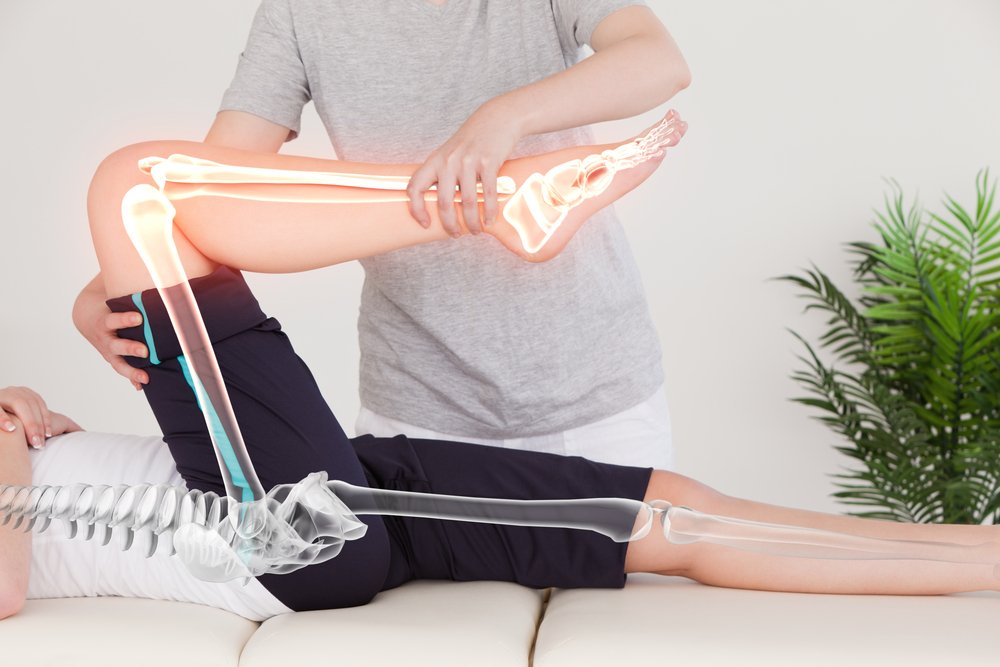
Rehabilitation after arthrodesis
After the operation, you will remain in the center's inpatient department for 1-3 days and will be monitored by medical staff around the clock. On the 12th to 14th day (between bandages, under constant observation) you must come to the clinic to have the stitches removed. You will be x-rayed monthly for 3-4 months and monitored by our trauma surgeon. If you have leg joint surgery, you may only be able to walk with crutches for the first two months. The next phase of rehabilitation is to select and perform physiotherapy exercises and develop general mobility. Depending on the joint being operated on, this process can take three months or longer.
Arthrodesis in the surgical center 'SM-Clinics' offers highly qualified doctors, modern methods and cost-effective treatment. Call us at +7 (495) 292-59-87 to learn more about our services and to schedule an appointment with a trauma surgeon.
Read more:- How much does knee surgery cost?.
- Anatomy of the subtalar joint.
- dislocation of the ankle.
- metatarsophalangeal joint.
- Ankle ligament sprain.
- Schopar and Lisfranca joints are.
- pelvic thigh joint.
- Shapar joint.