As a rule, surgical intervention is necessary only in particularly difficult situations: when a purulent or necrotic process has developed, when there is a significant deformity of the foot or when the tissue is severely damaged. In the other cases, a conservative treatment plan is applied based on:
- What is a hollow foot?
- Causes of hollow foot.
- ICD-10
- causes
- causes
- Foot anatomy
- orthoses
- Severity of flat feet in adults
- Types of flat feet in adults
- Longitudinal flatfoot
- Transverse flatfoot
- Fixed flat foot
- Unfixed flatfoot
- The high arch of the foot and everything that goes with it
- What causes this?
- What are the risks?
- What can I do if my feet have high arches?
- Possible causes of pain
- Anomalies in which the arch of the foot hurts on the inside
- opinions of doctors
- Soft tissue surgery
- Release of the plantar fascia (Steindler operation)
- Transferring the long fibular tendon to the short fibular tendon
- Lateralization of the tendon of the tibialis anterior muscle
- Corrections of toe claw deformities
- Bariatric Surgery
- Osteotomies
- arthrodesis
- Video about our clinic for traumatology and orthopedics
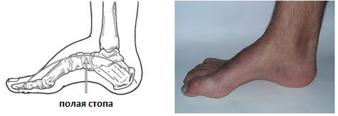
What is a hollow foot?
A hollow foot is characterized by a high instep and arch, resulting in abnormal load distribution, pain and instability. Cavus foot is most commonly associated with neurological disorders, can occur at any age, and can affect one or both feet. One of the most common hereditary causes of hollow foot is Charcot-Marie-Toute disease.
Causes of hollow foot.
The formation of hollow feet is most commonly associated with neurological diseases such as cerebral palsy, Charcot-Marie-Toute disease, polio, muscular dystrophy, sequelae of a stroke, or spina bifida. However, in some cases, isolated hollow foot is identified as a variant of an inherited structural anomaly. A correct diagnosis is important because it can predict the course of the disease and the prognosis. If the deformity is caused by a neurological disorder, it will be stable. However, if cavus foot is an isolated structural anomaly, it usually does not progress any further.
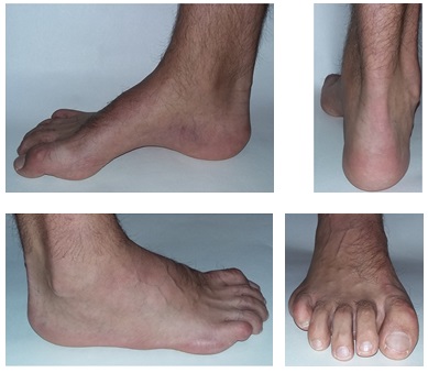
The arch of the foot and the instep of the hollow foot are more pronounced than normal. In addition to the high arch and instep, the following features may be present:
-Hammer toes or claw-shaped toe deformity.
-Calluses (hyperkeratosis) on the toes and feet due to abnormal load distribution.
-Pain in the foot when standing and walking, which is more noticeable on the outside of the foot.
-Instability of the ankle joint, primarily due to varus inclination of the heel bone.
Some people with a hollow foot suffer from what is known as 'cave foot', which is due to muscle weakness in the lower leg and a disturbed muscle balance. This always indicates a neurological cause of the hollow foot.
ICD-10
A cavus foot is an excessive enlargement of the arch of the foot. It can occur in a variety of diseases of the nervous system and musculoskeletal system. It can occur after severe trauma to the foot (bruises, severe tarsal bone fractures), especially in childhood. Sometimes inherited. Associated with rapid fatigue and pain when walking. Causes blisters and deformation of the toes. In some cases there is no functional impairment.
The reason for treatment is usually severe pain in the feet and the inability to wear appropriate footwear. Conservative treatment is carried out for mild or moderate hollow feet. If the deformity progresses, surgery is indicated. Orthopedists/traumatologists are involved in the treatment. If the pathology is caused by a disease of the nervous system, neurologists simultaneously treat the underlying disease.
causes
The exact mechanism for the development of pes cavus has not yet been clarified. It is believed that the pathology is most commonly due to muscular imbalance resulting from hypertonicity or paretic weakness of certain muscle groups of the lower leg and foot. However, experts note that when examining patients with pes cavus in some cases, it is not possible to detect a clear increase or decrease in muscle tone.
Cavus foot can occur in a number of diseases and neuromuscular malformations, including. in poliomyelitis, muscular dystrophy, spinal dysraphia (incomplete closure of the medial spinal suture), Charcot-Marie-Tooth disease (hereditary sensorimotor neuropathy), polyneuropathy, syringomyelia, cerebral palsy, Friedreich's ataxia (hereditary ataxia due to damage to the spinal cord and cerebellum), Meningitis, encephalitis, malignant and benign spinal cord tumors. In rare cases, the pathology is caused by burns on the foot or improperly healed fractures of the heel and calcaneus. In about 20 % cases, the triggering factors for the deformity remain unknown.
causes
In about 3 % cases, the disease is congenital and is based on intrauterine malformations of the bones and ligaments of the foot. Trauma, including fractures of the tarsal and metatarsal bones and ankle fractures, can also be a cause of the condition. There is also paralytic flat foot, which is caused by paralysis or paresis of the foot and lower leg muscles, and wobbly flat foot, which is caused by deformation of the bones due to excessive softness.
However, the most common condition is static longitudinal flatfoot, which is caused by weakness of the ligamentous and muscular apparatus of the distal parts of the lower limbs. Factors that favor the development of static flat feet include obesity, pregnancy, excessive physical activity, jobs that require long periods of standing (salespeople, receptionists, wood turners, etc.), wearing uncomfortable, poor-quality footwear and weakening of the ligaments and Muscles of the foot due to age or lack of physical activity.
Foot anatomy
The human foot consists of 26 bones, numerous joints, ligaments and muscles. All of these anatomical elements are connected to each other and form a unit that provides support and walking functions. The correct function of the feet enables optimal distribution of body weight when moving in space and ensures correct posture and physiological alignment of the foot, knee and hip joints. When walking, the foot absorbs ground shock, thereby reducing the load on the upper parts of the musculoskeletal system.
The interconnected components form two longitudinal arches in the foot. The longitudinal arch is located at the outer edge of the foot and the transverse arch is located at the base of the toes. Due to this complex arch shape, the foot does not touch the base with the entire sole, but only with certain points: on the heel bone, at the base of the 1st and 5th toes. With a longitudinal arch, the height of the longitudinal arch is lowered and the foot touches the base with almost its entire surface.
This leads to inadequate load distribution, deterioration of the cushioning properties of the foot, incorrect posture and the development of abnormal movement patterns. As a result, progressive pathological changes develop not only in the feet, but also in other parts of the musculoskeletal system. This increases the likelihood of coxarthrosis, gonarthrosis, osteochondrosis and other degenerative-dystrophic diseases.
orthoses
Orthotics for people with high arches are custom-made. They have a special design that effectively supports the foot while walking and distributes pressure evenly. They ensure pain-free and comfortable movement and prevent further deformation of the hollow foot.
Custom-made insoles do not need to be adjusted and have a long lifespan (approx. 2-3 years).
The shape, height and stiffness of the orthoses are selected individually. This makes their use sensible and safe.
Hollow feet can be easily corrected. The most important thing is to see a doctor as early as possible. Appropriate therapy and the use of special insoles help to control high arches and eliminate pain when walking.
Severity of flat feet in adults
Each form of flat foot has its own characteristics, so doctors usually consider the degree of deformity for longitudinal and transverse forms separately.
Depending on the severity of the pathology, orthopedists classify the IV degrees of flat foot:
| Grade I | mild, almost symptomless, fatigue and pain in the legs sometimes at the end of the day; easily correctable |
| Grade II | pronounced and painful feet, ankles and calves, swelling and heaviness of the feet at the end of the day, the gait may be altered and the foot deformity is already visible externally. |
| Grade III | Severe foot deformity - almost no arch, constant pain in the lower leg, knees, hips and lower back. Against this background, spinal curvatures, joint wear and osteochondrosis, herniated discs and headaches can develop. The appearance of a crunch in the knees means that the joints have begun to disintegrate. If left untreated, this stage can lead to disability. |
| Stage IV | Inward rotation of the sole, severe pain, limitation of movement, deformation of the entire skeleton |

Types of flat feet in adults
Depending on which arch of the foot is affected by the deformity, flat feet can be longitudinal or transverse and immobile or non-mobile.
Longitudinal flatfoot
The inner longitudinal arch of the foot deforms so that the sole of the foot comes into almost complete contact with the ground and increases in length. If severe, toe splaying and an X-shaped structure may develop. Signs of fatigue and pain in the foot can occur even with moderate symptoms.
If the longitudinal arch is deformed, the foot deviates from its central axis and collapses inwards, this is called a valgus flatfoot.
The following are more likely to be affected by this form of flat foot:
- elderly people;
- Athlete;
- hairdressers and painters;
- pregnant women;
- women wearing high heels;
- sedentary and obese people;
- People with leg injuries.
Transverse flatfoot
The forefoot is deformed and the big toe deviates outwards. This leads to a bulging of the transverse arch. Patients develop calluses and corns on the soles of the feet and the foot contracts. In addition to the thumb, the second and third toes are also deformed. They appear crooked and the curvature increases with the bulge of the bump on the big toe - the valgus bone.
Due to the changed pressure points, the foot becomes wider and those affected have difficulty fitting into shoes. Patients also complain of pain in the metatarsophalangeal joints of the toes. This form of flat foot occurs most often in women between the ages of 35 and 50.

Fixed flat foot
The degree of deformation of the arch of the foot does not change with the load on the foot.
Unfixed flatfoot
The height of the arch of the foot decreases as the load on the foot increases.
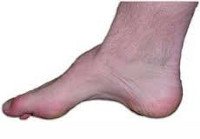
The high arch of the foot and everything that goes with it
The human foot can have a normal, low or high arch. The first variant is not associated with abnormalities, pain or gait disorders. In the second case, a low arch means that the person suffers from a disease - flat feet. It causes pain, swelling, and gait disturbances that can lead to valgus foot deformity and ingrown toenails.
A high arch of the foot is one of the abnormalities, as well as flat feet. If the arch of the foot is high, the ability of the foot to rotate is significantly limited. The foot sinks towards the outer edge and the load is distributed unevenly from the big toe to the little toe and the ring toe.
What causes this?
You can see the elevated type of athlete's foot with the naked eye. If you place the foot on a flat surface, you will notice that the middle part of the arch of the foot rises above the surface. This is often seen in ballerinas. It may be a result of muscle hypertonicity caused by certain neurological diseases. Spinal cord tumors, cerebral palsy, polyneuropathy and poliomyelitis can also promote the development of the pathology.
What are the risks?
The disadvantages of an arched foot are quite numerous. For example, the foot may not fit into the shoe but rest on the toe of the shoe. Women may experience discomfort or even pain when wearing high-heeled shoes or sandals.
People with this peculiarity have a significantly disturbed gait and increased pressure on the ankles. Calluses form on the soles of the feet and the feet shrink. The toes are particularly affected, as they become deformed and clawed over time. In severe cases, a person wearing high heels can become physically disabled and partially unable to work.
What can I do if my feet have high arches?
In children, foot pathology is detected during medical examinations. In adults, the arch of the foot is too high, which can lead to gait problems or pain when walking.
An accurate diagnosis can only be made by a doctor. If problems are identified, various physiological treatments and massages may be prescribed. Wearing special orthopedic shoes and insoles plays its own role in correcting arch deformities.
Vibrating orthopedic insoles have a positive effect on such pathologies. They are considered to be the salvation of people with high arches as they eliminate all the discomfort associated with it. They can be worn by anyone. They are used for both therapy and prevention.
If you think that the information on this page will be useful to your friends, acquaintances and colleagues, you can share it on your social network. To do this, simply click on the corresponding symbol:
Possible causes of pain
Attempting to relieve arch pain from the inside often only makes the condition worse. To eliminate this unpleasant symptom, the cause of the pain must be correctly identified, which can only be done by a doctor.
- Inflammatory diseases;
- mechanical trauma;
- Weakening of the ligaments or joints;
- degenerative and dystrophic pathologies;
- various types of foot deformities;
- autoimmune abnormalities;
- metabolic disorders;
- long-term trauma;
- Other diseases.
The appearance or worsening of arch pain on the inside of the foot is often triggered by the following factors:
- hypothermia (including wearing inappropriate, lightweight footwear);
- uncomfortable footwear;
- Excessive exposure to sporting activities;
- Hormonal imbalances;
- Obesity;
- hypodynamia;
- injuries;
- infections;
- unbalanced diet (including weight loss diets);
- Persistent fatigue; Lack of sleep;
- dehydration;
- Exacerbation of chronic diseases.
If you are affected by one of these diseases or have recently been affected, you should definitely talk to your doctor about it.
Anomalies in which the arch of the foot hurts on the inside
Rapid foot fatigue and foot pain are common in clubfoot, flatfoot and similar conditions. Wearing high-heeled shoes and shoes with flat soles increases the risk of foot pain.
After the age of 30, the risk of osteoarthritis increases. It is a disease that leads to the progressive destruction of cartilage tissue, including in the ankle and toe areas. A slight discomfort eventually develops into severe pain. The pain gradually gets worse:
Arthritis is an equally dangerous disease. It is accompanied by persistent inflammation (infectious or non-infectious) that attacks the cartilage. In infectious arthritis, the inflammatory process develops rapidly and leads to severe swelling and deformation of the foot, as well as fever. This form of arthritis is usually preceded by another illness - an acute respiratory infection, flu, purulent sore throat or food poisoning.
Gout, a disease caused by an imbalance in purine metabolism, can also cause pain on the inside of the foot. Gout often causes tophi – lumps with a thick, whitish fluid inside. Uric acid crystals build up around the joint, injuring the tissue, causing swelling and inflammation, and deforming the foot. It is not uncommon for gout to cause pain in the ball of the foot on the inside of the foot. Gout is also characterized by severe, sudden pain attacks (usually at night), which are accompanied by swelling and redness of the skin. After a certain period of time the pain subsides spontaneously, but without treatment the attacks become more frequent.
The inside of the foot can hurt due to inflammatory conditions such as ligament and tendonitis. In the former, the ligaments are affected, in the latter, the tendons. Both conditions can be caused by wearing uncomfortable shoes, tissue immobility, trauma, chronic illness (especially diabetes), and obesity.
opinions of doctors
These are real people's opinions, taken from the doctor's website on prodoctorov.ru
My name is Elena Vorobyeva. In August 2021 I had hip surgery. In February 2022 I had another check-up. The doctor said everything was fine.
I went to Denis Sergeyevich to get advice about knee arthroplasty. Gonarthrosis had previously been diagnosed in the same clinic and surgery was recommended. Doctor.
My grandmother (91 years old) had a complicated fracture of her upper arm. Nobody wanted to help her, and it was scary to send my grandmother to the hospital with Kovds. Several doctors said they couldn't help her. from.
I, Mitropolevsky Tatiana Valentinovna, born in 1949, underwent surgery for cemented total endoprosthesis of the left hip joint. The first similar operation was on the right joint.
I live in the Rostov region. For the last two years I was very worried about the pain in my hip joint, nothing was working anymore and I decided to have an endoprosthesis inserted. On the advice of a surgeon in Rostov, I decided to have the operation.
For over a year I had recurring pain in my pelvic area that got worse when sitting or lying on my side. I went through all the doctors: gynecology, urology, proctology, neurology, etc.
I went to the doctor with severe pain in my hip joint and I could hardly walk. Denis Sergeevich examined me thoroughly, prescribed an examination, after which manipulations were carried out.
dr Yakushev Denis Sergeevich is an orthopedist and traumatologist, but he is also a doctor and a man of stature. dr Yakushev operated on my mother's right hip joint in 2016. The operation was an emergency.
I really liked everything about it! The doctor thoroughly explained everything and explained the situation. Clear and understandable! He was very accommodating and professional! It was a pleasure speaking with him. I recommend him to 100%!!!
I would like to thank Denis Yakushev very much! I went to him for a torn bicep tendon. The diagnosis was made very quickly and an operation was scheduled immediately. The operation was carried out by.
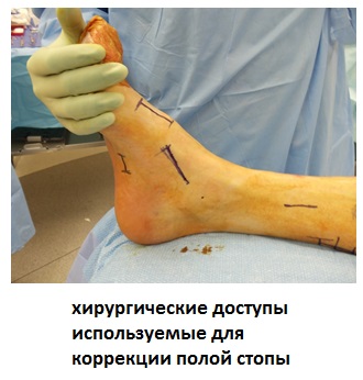
Soft tissue surgery
Release of the plantar fascia (Steindler operation)
The reduced plantar fascia plays a very important role in maintaining the excessive height of the longitudinal arch in patients with cavus foot deformity. By relieving the longitudinal fascia, the height of the arch of the foot can often be reduced if the patient still has some mobility in the deformity. Sometimes this operation can be performed in combination with lateralization of the calcaneus, triquetral arthrodesis, or first metatarsal osteotomy.
During the operation, the fascia is stretched and crossed where it attaches to the heel bone.
Transferring the long fibular tendon to the short fibular tendon
In many cases of cavus foot deformity, particularly in patients with Charcot-Marie-Tooth disease, there is paralysis of the tibialis anterior muscle, but function of the long fibula is preserved. The latter collapses and deforms the forefoot, resulting in cavus foot deformity. This can be fundamentally changed by lateralizing the long fibula tendon to the short fibula, thereby restoring the normally lost function of the short fibula.
Lateralization of the tendon of the tibialis anterior muscle
Shifting the insertion point of the tibialis anterior tendon to the lateral tibial spine is a very effective tool for changing the biomechanics of the foot while maintaining adequate muscle strength. It enables increased stability of the rear foot and shifts the center of force of the ankle joint outwards. The operation carries minimal risk of overcorrection and is technically simpler than other procedures.
Corrections of toe claw deformities
Claw deformities of the toes are common associated deformities of the cavus foot. The claw deformity often resolves itself by correcting the metatarsal and hindfoot position.
Bariatric Surgery
Osteotomies
Sometimes a deformity of the bone involved in the formation of hollow foot affects the patient's ability to put normal weight on the foot, while leaving other parts of the foot relatively unaffected. This is usually a fixed varus deformity of the hindfoot or a fixed plantar flexion of the first ray. In some cases, it is possible to correct a fixed deformity and straighten the foot again without arthrodesis (artificial joint closure).
Osteotomies of the first metatarsal or calcaneus or both are usually performed in conjunction with soft tissue surgery (releasing the plantar fascia and transferring the long fibular tendon to the short fibular tendon).
arthrodesis
The three-joint arthrodesis is indicated for all more or less severe, fixed foot deformities. In patients with a cavus foot deformity, in addition to the three-joint arthrodesis, other corrective procedures may also be performed, such as osteotomy of the first metatarsal bone or the heel bone.
Correction of the heel bone through a three-joint arthrodesis restores the normal support function of the foot.
The analysis of the results of the various surgical options for the treatment of hollow foot suggests a significant improvement in foot function, pain relief and good to excellent treatment results in 70 percent of patients, even in the long term. Patients with cavus foot deformity and pre-existing osteoarthritis have poorer treatment outcomes.
Foot deformity may recur to some extent after surgery, but most patients can continue to wear regular footwear and do not need to wear orthotics all the time.
Video about our clinic for traumatology and orthopedics
Read more:- Treatment of the hollow foot.
- Equinos pes cavus.
- The formation of the arch of the foot.
- Excessive pronation of the foot.
- heel bone injury.
- dislocation of the foot.
- Anatomy of the heel bone x-ray.
- polo varus.