Grade I is the most effective treatment for a patient with early activation of the injured limb.

- Torn ligaments in the ankle joint: symptoms
- Ligament groups of the ankle joint
- Types of Ankle Injuries
- Consequences and complications of ankle injuries
- diagnosis
- Different types of ruptures
- Damage to the hip joint-clavicle junction
- Ligament injuries in the ankle joint
- Damage to the collateral ligament of the big toe
- Heel tendon injury
- Extent of ligament injury
- Surgery for a torn ligament
- rehabilitation
- 3. Ankle sprain treatment
- Symptoms of a tear of the anterior cruciate ligament
- Symptoms of a rupture
- Indications for an ultrasound examination of the ankle joint
- How does the ultrasound examination of the ankle work?
- pain in the knee joint
Torn ligaments in the ankle joint: symptoms

Ankle ligaments are tendon ligaments whose main function is to stabilize the joint. They stabilize the bones and prevent them from moving apart.
The tendon ligaments are strong, but not flexible, which is why the ligaments of the ankle joint can tear: partially or completely, but not dislocated, as this pathology is incorrectly called.
Ligament groups of the ankle joint
The ligaments that fix the ankle joint are divided into three groups:
- First group. The first group of ligaments includes the patellofemoral ligaments, the anterior patellofemoral ligaments, and the posterior patellofemoral ligaments, which run along the lateral malleolus. They prevent lateral displacement of the talus;
- second group is represented by the deltoid ligament (internal collateral ligament), which consists of two layers: a superficial one, adjacent to the ankle joint and the talus, and a deep one, adjacent to the talus from the inside;
- third group the intercondylar syndesmosis, the posterior transverse ligament and the intercondylar ligaments: posterior and anterior.
The anterior cruciate ligament is the most commonly injured ligament of the outer group, especially the anterior fibula in leg rolling, sports injuries, high jumps and falls. The risk groups for ankle injuries include women who wear high-heeled shoes for a long time, the elderly due to a weakened musculoskeletal system with degenerative changes and children with high mobility, people with high foot pressure and congenital foot deformities such as flat and club feet, and people with previous ankle injuries.

Types of Ankle Injuries
Now let's take a look at the main types of ankle injuries that can occur in patients of very different age groups. Each injury requires a specific first aid approach. Therefore, you should not try to diagnose and treat the injury yourself. If you have suffered a traumatic impact, you should see a trauma surgeon immediately. Only this specialist can make an accurate diagnosis, provide high-quality first aid and prescribe initial treatment.
The following are the main types of ankle injuries:
- Fracture or breakage of the condyles of the tibia, femur;
- fracture or break of the talus or heel bone;
- Sprain and strain of the ligaments and tendons;
- Sprain, strain and tear of the joint capsule;
- Hematomas in the surrounding soft tissues;
- Subluxations and dislocations.
Each of these injuries has its own peculiarity. Fractures and fractures almost always do not heal properly without expert medical care. A thickened bone callus forms, which puts pressure on the surrounding soft tissues. In this way, post-traumatic angiopathy and neuropathy can develop. In most cases, a cast is required to immobilize the injured limb for up to 40 days. Once the cast is removed, full rehabilitation at a chiropractic clinic is required.
Sprains and microscopic tears in ligaments and tendons are no less dangerous. It usually starts with a mild sprain of the foot. For example, you tripped or sprained your foot. A small tear occurs in a single fiber.
Without professional help, the exact same ligament or tendon tissue will not form again at this point in the healing process. This is because tendons and ligaments do not have their own blood supply. They receive fluid and nutrients only through diffusion exchange with the surrounding muscles. To do this, however, the muscles have to work constantly.
Consequences and complications of ankle injuries
Without proper treatment, various complications arise after an ankle injury. Some of these lead to disabilities or severely limit mobility.
For example, a contracture of the ankle joint after an injury means a complete loss of mobility, which makes walking very difficult. But that is not all. If contractures occur in the ankle joint, the distribution of the damping forces is disturbed. The knee and hip joints begin to deteriorate rapidly. Deforming gonarthroses and coxarthroses arise. Within 2-3 years the lumbosacral spine is destroyed.
Other possible consequences of an ankle injury include:
- Destruction of the small joints of the foot bones;
- Development of flat feet or club feet, which have a negative impact on the entire musculoskeletal system;
- Stiffening of the joint (ankylosis) with subsequent progression to complete or partial contracture;
- Deformative arthrosis of the ankle joint – thickened bony outgrowths form on the bone surfaces, which hinder movement and cause severe pain;
- Rupture or complete collapse of the ligaments and tendons;
- Hemarthrosis, which is caused by blood entering the joint capsule;
- Aseptic inflammatory necrosis of bone and cartilage.
All consequences of a traumatic impact can be easily prevented. First of all, all possible risk factors should be eliminated from your life. Ankle injuries most often occur in people who have one or more of the following risk factors:
- Excessive body weight - the higher the weight, the more stress is placed on the tissues of this joint, increasing the likelihood of suffering a serious injury in the event of a slip or sprain;
- Sedentary lifestyle - the lack of regular physical activity weakens the muscular skeleton and leads to dystrophic and atrophic degeneration of the joint, which thereby loses its ability to withstand greater mechanical stress;
- poor diet and inadequate intake of clean drinking water throughout the day;
- Smoking and drinking alcoholic beverages
- Hard physical work;
- unsuitable footwear for everyday use and sports;
- poor foot position in the form of clubfoot or flatfoot.
diagnosis
The diagnosis is made on the basis of an examination and stress tests. Sometimes additional tests (instrumental and laboratory) are performed to detect damage to other structures. Before treating a torn meniscus ligament, it is important to ensure that the meniscus itself is not damaged.
Stress tests place stress on the ligaments and tendons - such tests help determine which specific components are damaged. The test requires the patient to move the joint in the opposite direction to its natural direction.
Due to the acute pain during the test, muscle spasms may occur, which does not allow detecting an unstable part of the limb. To avoid inaccurate results, wait until the muscles are as relaxed as possible and perform the exam again. If the muscles are tense, anesthesia should be applied and the examination repeated. If damage to several groups of ligaments is suspected, all ligaments and tendons should be examined. The diagnosis and treatment of a knee ligament rupture can be combined with the diagnosis and treatment of an Achilles tendon rupture.
Simultaneously with the examination of the injured limb, the healthy limb is loaded and the results are compared:
- In grade I fractures, joint function is the same, but functional loading is painful;
- Grade II ruptures are painful and joint function is limited;
- Grade III ruptures are less painful. This is because the ligaments or tendons are completely torn and do not deform during examination.
Different types of ruptures
How long does rehabilitation take after a ligament or tendon rupture in the foot or hand? That depends on the localization. A rough prognosis can be made based on the severity of the symptoms.
Different ligaments and tendons show different signs of tearing.
Damage to the hip joint-clavicle junction
Caused by a fall from the shoulder rest or abduction of the shoulder.
If the acromioclavicular ligament is severely torn, the clavicle may shift forward in relation to the hip socket.
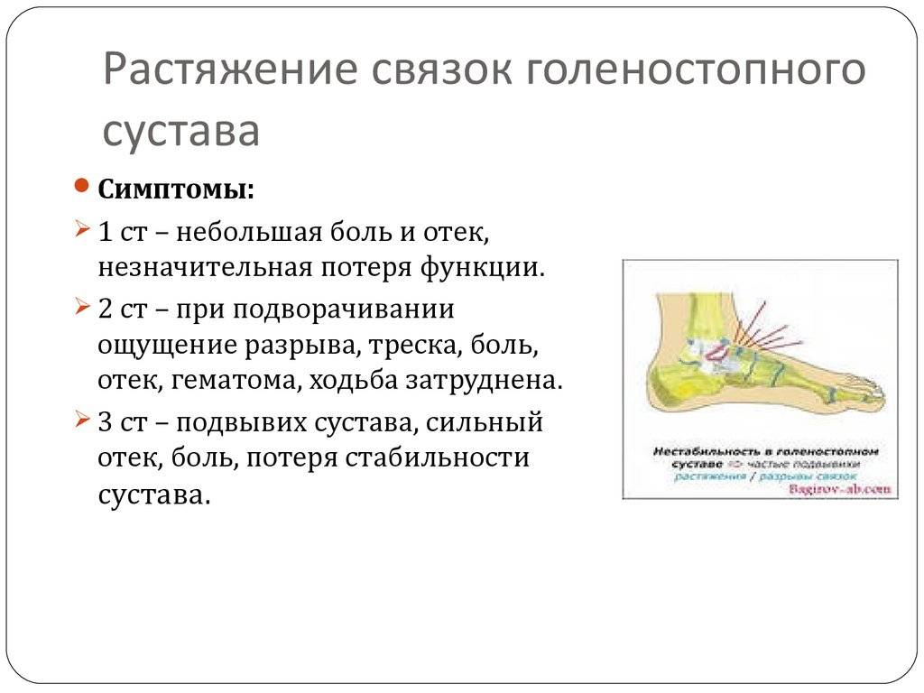
Ligament injuries in the ankle joint
This is a very common type of injury. It most commonly occurs when the foot is turned inward. It can also be injured:
Severe tears (Grades II and III) can lead to a chronic ankle sprain. With a grade III tear, the swelling causes the joint to look like a large egg. The swelling and pain are more pronounced on the anterolateral surface of the joint. The recovery time from a torn ankle ligament may depend on which ligament was damaged.
Damage to the collateral ligament of the big toe
What can be observed is the so-called extensor finger - the abduction of the toe. During the stress test, the toe is abducted in the direction of the radius.
Heel tendon injury
The stress test consists of dorsiflexion of the foot. Passive soleus flexion of the foot is impaired when the patient's calf is compressed. Partial cracks may not be detected.
Extent of ligament injury
At this point in the article a legitimate question arises: What treatment is appropriate for an ankle ligament injury?
FirstFirstly, each patient must be treated individually, as the degree of ligament damage, swelling and pain syndrome must be assessed and perhaps even further investigations carried out.
Secondly:First: Treatment should be recommended according to the findings described in the first point.
- cold
- Kinesio taping
- soft or rigid orthoses
- Prescription of anti-inflammatory medications
- topical medications
For more in-depth advice, you can contact our clinic at any time. We have professional doctors who will make the correct diagnosis and prescribe treatment until complete recovery.
Surgery for a torn ligament
The choice of treatment for a torn ligament depends on the extent of the injury, the severity of the symptoms, the bone or muscle injury, etc. If no more than 50 % of fibers are damaged, conservative treatment is sufficient: immobilization, painkillers and exercise during the recovery period.
If the damage is more severe, healing can only be achieved through surgery. The arthroscopic operation is carried out via a 'closed' approach through several punctures into the joint protrusion. It is indicated for long-term injuries or tears of ligaments within the joint. This method is most often used for knee and anterior cruciate ligament injuries.
The essence of the procedure is to reconstruct the damaged ligament through the implantation of autografts (using the patient's own donor tissue) or, less often, allografts (synthetic or biological prostheses from a donor bank). Self-absorbing screws are used for fixation. The operation can restore the stability and function of the joint.
Special types of sutures and various fixation devices can be used in the surgical treatment of fresh ligament tears. The choice of surgical technique is made by the treating traumatologist based on the individual clinical picture, age, physical activity and lifestyle of the patient.
In most cases, timely surgery allows for complete recovery of the joint and even a return to high levels of physical activity.
Traumatologists have a number of methods for treating ligament injuries, but their effectiveness largely depends on the timing of treatment. Attempting to repair a torn ligament on your own can lead to unpleasant consequences: inflammation, deformation of the limb and instability of the joint with an increased risk of re-injury.

rehabilitation
The duration and type of postoperative rehabilitation depends on the type of joint, the extent of the injury and the extent of the surgery. You will usually be discharged from the hospital one to two days after the operation. After that, you must take the prescribed medication and follow your doctor's instructions regarding your exercise habits.
If you have ligament damage in the lower limbs, you should use crutches and gradually increase the load on the affected leg. In consultation with the doctor, the flexion angle of the joint is also increased. The final phase of postoperative rehabilitation includes special physical therapy.
The symptoms of ligament damage are very similar to those of a joint fracture, so an accurate diagnosis can only be made after a thorough examination.
Physiotherapy will significantly speed up the healing process. If your doctor has recommended a specific course of rehabilitation, you should not ignore this recommendation.
'Emergency Medical Care', edited by JE Tintinalli, Rl. Croom, E. Ruiz, translated from English by VI Kandror, MV Neverova, MD, AV Suchkov, MD, AV Nizov, JL Amchenkov; ed. by VT Ivashkin, MD, PG Brusov; Moscow 'Medicine' 2001.
Traumatology and orthopedics: a textbook / edited by NV Kornilov. – Edition 3, revised and updated – M.: GEOTAR-Media, 2016.
3. Ankle sprain treatment
It is important to follow established procedures It is important to follow the first steps after the injury. However, if pain and other symptoms persist or even worsen 2-3 days after spraining your ankle, you should definitely see a doctor.
In severe cases of sprains For severe ankle sprains, the doctor will apply plasters or special stabilizers. In rare cases, the patient may even need surgery. In such cases, rehabilitation is necessary because prolonged immobilization of the joints leads to a weakening of the muscles of the ankle joint. Prolonged immobilization of the joints leads to a weakening of the muscles and cartilage structure, sensory disturbances and contractures that limit mobility. The treatment usually lasts several weeks .
If the joints were injured, you should use it for 2-3 weeks maintain a diet rich in collagen, the most important component of connective tissue. To maintain its production, eat foods that contain the amino acids that contribute most to collagen proteins collagen proteins. These include beetroot and other red vegetables (silicon), Onions, leeks (sulphur), buckwheat groats, figs, apricots..
You also need lots of vitamin C, which is essential for the formation of collagen. Good sources for this are e.g. B, Citrus fruits, parsley, red pepper. You should also just buy traditional meat (without artificial additives that can have an anti-inflammatory effect, and the high sodium content makes swelling last).
Symptoms of a tear of the anterior cruciate ligament
Most people hear a cracking sound when they injure their knee and notice the development of the following symptoms:
- A pain syndrome that increases as the swelling of the knee joint increases
- Swelling of the joint and surrounding tissues, which increases in the first 24 hours after the injury
- Restricted or excessive joint mobility
- Uncertainty, insecurity, 'weakness' of the knee joint
Symptoms of a rupture
Reliable signs of a cruciate ligament rupture are the 'anterior and posterior drawer' symptoms. To detect these, the patient lies supine on the table and the doctor sits at the patient's feet so that the patient's foot rests on the thigh. The doctor places their hand on the top third of the tibia and tries to move it backwards or forwards. If the tibia moves backwards or forwards excessively, the test is positive.
The anterior cruciate ligament is classified as follows:
- Complete – with damage to all fibers of the cruciate ligament. Characterized by severe pain syndrome, massive swelling and loss of joint stability
- Tear of the cruciate ligament from its attachment point on the femur or tibia. It is also characterized by a severe pain syndrome, massive swelling and a loss of stability of the joint.
Indications for an ultrasound examination of the ankle joint
In addition to obvious injuries, ankle problems, pain of varying severity or type, and restricted mobility can also be reasons for an ultrasound examination. To understand what is the cause of these symptoms, the doctor needs to know exactly the condition of the joint.
The examination is recommended in the following cases:
- for bruises, injuries, any injury in this area;
- in cases of misalignment
- if degenerative processes such as arthrosis, arthritis are suspected;
- pain of varying intensity and nature;
- restriction of mobility of the joint;
- if the ankle is swollen, red and hot;
- before a surgical procedure;
- after surgery to determine the results and dynamics of recovery;
- In the presence of a heel spur;
- for foot deformities and flat feet.
Ankle joint problems are also associated with systemic diseases such as diabetes, problems with the musculoskeletal system and spine, and neurological disorders.
If the patient plays sports or dances - whether as a professional or as an amateur - torn ligaments and tendons can be the cause of the pain.
It is important to know that inflammatory processes do not cause symptoms in their initial stages. Since therapy is particularly effective at this time, active people, the elderly and people who put a lot of strain on their joints should undergo a preventive ultrasound examination of the ankle joint.
You do not need a referral from your doctor for the examination. You can do an ultrasound scan on your own and then contact a specialist.
How does the ultrasound examination of the ankle work?
During the diagnosis, which takes about 20 minutes, the patient sits or lies on a couch. First, socks, tights and pants must be removed to gain access to the joint.
A gel is applied to the skin in the area to be examined, then the diagnostician examines the ankle joint with a scanner. During the procedure, the doctor asks the patient to change positions several times to see the joint in different projections' – anterior, medial, posterior and lateral approaches.
In addition to the standard protocol, a Doppler blood flow study is usually also performed using a special attachment built into the ultrasound probe.
pain in the knee joint


Knee joint pain can have many causes, but before we talk about it, we need to understand how the knee joint works.
The knee joint is not only the largest joint in the human body, but also the most complex. It consists of three bones: the thigh bone (femur) at the top, the shinbone (tibia) at the bottom, and the kneecap (patella) in front of these bones. Both the femur and tibia have two protruding bony projections, the external and internal condyles. The outer condyle is called the lateral condyle (from the Latin word lateralis, outside) and the inner condyle is called the medial condyle (from the Latin word medialis, inside). The main movement of the knee joint is flexion, with the kneecap resting in the hip socket between the outer and inner condyles of the thigh.

Between the femur and tibia bones are the menisci - crescent-shaped layers of cartilage that increase the stability of the joint by increasing the contact area of the bones, acting as shock absorbers, and performing several other important functions, which you can learn more about in the article about meniscus tears.

The outer part of the joint is covered by the capsule. The inner layer of the capsule is called the synovium. The knee joint contains synovial fluid that lubricates the cartilage so it can slide better. The synovial fluid also supplies the cartilage with nutrients.
The ligaments in the knee joint ensure the stability of the bones in relation to each other. The most important ligaments of the knee joint are the anterior and posterior cruciate ligaments, the tibial collateral ligament (internal collateral ligament) and the medial collateral ligament (external collateral ligament).
Read more:- Anterior shin muscle (tibialis anterior).
- Treatment of torn ligaments in the ankle.
- Ankle ligament strain, ICD.
- Injury to the ligaments of the ankle.
- Ligament damage in the right ankle.
- The hock is the place where.
- ligaments in the ankle.
- Broken ankle.