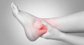
However, orthopedist Valentin Dikul claims that there is actually an effective remedy for joint pain! >>>
- Ankle capsule
- Innervation of the ankle joint
- Indications for ankle arthroplasty
- When is ankle surgery not possible?
- Indications and contraindications for use
- Types of immobilization devices
- elastic
- Radiological diagnosis
- How to choose the right endoprosthesis?
- preparation phase
- Joint injections for ankle arthrosis patient reviews
- Treatment of lateral epicondylitis of the elbow consists of
- What is needed for articular cartilage?
- Pain-relieving and anti-inflammatory tablets of the joint
- MRI in the SM Clinic
- Indications for MRI of the ankle joint
- TREATMENT COSTS
- FEEDBACK FROM OUR PATIENTS
Ankle capsule
The joint capsule of the ankle joint attaches to the cartilaginous edge of the articular surfaces and covers the front part of the ankle neck. The collateral ligaments are located on the sides of the joint and run from the ankle joint to the adjacent tarsal bones. Medial collateral ligamentThe medial collateral ligament (ligamentum deltoideum) has a plate that resembles the Greek letter delta. The lateral collateral ligament, lateral collateral ligament, consists of three bundles that extend from the lateral ankle in three different directions: anterior anterior talofibular ligament, inferior calcaneofibular ligament and posterior posterior talofibular ligament. The lateral ligaments reinforce the joint capsule and the capsule is thin anteriorly and posteriorly.
The ankle capsule is thin anteriorly and posteriorly. ankle capsule The flexor tendon of the long toe attaches posteriorly and the extensor tendon anteriorly.
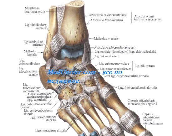
Innervation of the ankle joint
The ankle joint is innervated externally by the sural nerve, medially by the saphenous nerve and anteriorly by the deep peroneal nerve.

The information contained on this website is subject to the advice of a medical professional and is not a substitute for direct consultation with a medical professional.
Please refer to the User Agreement for further details.
Indications for ankle arthroplasty
In order not to miss dangerous damage to the bones and joints of the foot, which can lead to disability, it is important to pay attention to symptoms such as stiffness and pain. Timely surgical intervention can restore natural foot movement and a healthy gait. This avoids negative effects on the knee and hip joints and other parts of the human musculoskeletal system. The ankle joint is formed by the articular surfaces of the talus, tibia and fibula and is exposed to significant dynamic and static loads.
- Pathological processes in the tissues of the joint – osteoarthritis of various types (post-traumatic, age-related, dystrophic). Metabolic disorders, flat feet, rheumatism, gout, psoriasis, trauma and sprains can lead to osteoarthritis of the ankle;
- rheumatoid arthritis, an inflammatory disease that destroys the connective tissue of the joints;
- Ankylosis (immobility) or contracture (significant limitation of movement) of the joint as a result of injury, scarring or inflammation;
- Congenital anomalies of joint formation.

Sometimes bone hypertrophy and joint cysts are also an indication for surgery.
When deciding whether to insert an endoprosthesis, the doctor assesses the condition of the ankle joint itself and whether the patient has any concomitant diseases. The presence of valgus and varus deformities as well as flat feet is also taken into account.

When is ankle surgery not possible?
As already mentioned, the final decision on the possibility and advisability of endoprosthesis is made by the attending physician after examining the patient, reviewing his medical history and conducting clinical and radiographic examinations.
Contraindications to prosthetic surgery may include the following.
- Young age, when the skeleton is not yet fully formed,
- Osteoporosis, which prevents the prosthesis from being firmly anchored in the patient's bones
- Valgus and labral deformities of the foot with a large angle of deviation,
- Inflammatory processes in the joint area (the operation can be carried out after the inflammation has healed),
- Previous arthrodeses with nonunions,
- Soft tissue defects in the bones of the foot, damage to the ligaments if this means that the prosthesis cannot be properly adjusted and fixed,
- viral, bacterial and fungal infections as well as severe immune deficiencies.
Some contraindications are temporary, and if they are eliminated, the operation can still be performed.
Factors affecting postoperative recovery and effectiveness of arthroplasty include long-term glucocorticosteroid therapy, nicotine abuse, obesity, and excessive exercise. To increase the chances of successful arthroplasty and restore mobility to the joint, the patient may need to make lifestyle changes.
Indications and contraindications for use
During the treatment of ankle pathology, greater loading should be avoided. The use of an orthopedic device is necessary to keep the joint still, accelerate the healing of torn muscles and ligaments, and prevent the recurrence of joint diseases. The use of an ankle orthosis is therefore indicated in the following cases:
- inflammatory pathologies: juvenile, rheumatoid, gout, infectious arthritis;
- destructive and degenerative changes in the joint structures, e.g. B. Osteoarthritis;
- Achilles tendon injury – partial tear or complete separation of the tendon from its bony attachment;
- Synovitis, bursitis of various origins;
- hypermobility of the joints, leading to frequent dislocations and subluxations;
- Secondary joint instability due to flat feet, valgus deformity of the thumb, obesity;
- Paralysis, paresis caused by disorders of the cerebral circulation, e.g. stroke;
- Rickets, congenital anomalies requiring long-term correction of the joint, which contributes to its correct formation in children;
- the postoperative period, the return to active function of the ankle after endoprostheses, arthrodesis or other surgical procedures.
Contraindications to ankle orthoses are trophic ulcers, diabetic foot, open wounds of the ankle joint. For open fractures, a plaster cast is first applied to avoid stress on the joint. Wearing a plaster cast is recommended during the rehabilitation phase.
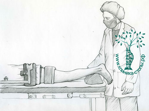
Types of immobilization devices
Domestic and foreign manufacturers produce various models of ankle braces for complete or partial immobilization. They differ in size, shape, purpose, construction, strength and price. Products that provide ankle support, ankle support, and/or ankle support are available at pharmacies and medical device stores. Your doctor may recommend a simple, soft bandage that does not restrict freedom of movement. However, sometimes an orthosis with a complex device with hinges, metal or plastic inserts is required.
elastic
This group includes ankle orthoses designed for sports and to prevent the recurrence of inflammatory and degenerative pathologies. They are designed to limit joint mobility, reduce increased muscle tension, eliminate pain and swelling. While wearing the product, light pressure is applied to the ankle joint, which has a warming and massaging effect. Elastic bandages are mass-produced, easily accessible and inexpensive. They are very easy to use: they are quick to put on, can be washed in warm soapy water and are easy to change into trousers, jeans or even tight tights. Especially for children, they come in bright colors and with different motifs to distract and cheer them up.
| Ankle supports | Purpose and therapeutic effect |
| Protective | Used for tissue repair after fractures. They have an antibacterial and disinfectant effect, prevent infections and prevent moisture from penetrating wounds. |
| Therapeutic | They resemble bandages made of elastic and solid gauze and are soaked in solutions of pharmacological agents. They are essential for accelerating metabolic and regeneration processes. |
| Compression bandages | Used to prevent and eliminate hemorrhoids. Their use allows you to quickly stop bleeding in joint cavities by applying gentle pressure to the blood vessels. |
| immobilization | Suitable for various injuries (severe bruises, partial tears of ligaments, muscles, tendons). Sports doctors recommend wearing insoles to relieve muscle pain after intense exercise or injury |
| Corrective | Suitable for the correct development of joints in children or to prevent further deformities in adults. Used for congenital and acquired defects to maintain the anatomically correct position of the ankle joint - valgus, hallux valgus foot |
Radiological diagnosis
A hypertrophied posterior talus, the os trigonum, is often seen on the x-ray. If there has been a previous trauma, we look for signs of an unhealed fracture in that area.
After a trauma, scintigraphy is often indicated if the x-rays show no changes. If the result is positive, a CT scan may be indicated, which is particularly helpful in post-traumatic cases when the extent of the injury and the location of calcifications or bone fragments need to be accurately assessed.
Conservative treatment of posterior epicondylitis syndrome includes restriction of movement, physical therapy (massage, stretching and muscle strengthening exercises), local ice packs, nonsteroidal anti-inflammatory drugs, and glucocorticoid injections.
For dancers, dance technique is analyzed and corrected, including correcting foot misalignments (flexion).
If conservative treatment is ineffective, intra- and extra-articular pathology of the ankle may require surgery.
Treatment includes removal of the hypertrophied posterior capsule of the ankle, os trigonum, hypertrophied posterior calcaneal process, false joint, calcifications, osteophytes and loose joint bodies.
With the development of minimally invasive surgery, new methods of treating this pathology have been developed, including endoscopic techniques, which have several objective advantages. The operation is performed on an outpatient basis and functional treatment begins immediately afterwards.
The operation is performed on an outpatient basis under general anesthesia or epidural anesthesia. The patient is placed on his stomach.
An arthroscope is inserted into the joint cavity through two skin incisions at the back of the ankle, and the images are displayed on screens in the operating room. We perform the surgery in an aqueous environment to improve joint alignment and flush out pathological tissue - for this we use a physiological solution or a special Ringer's solution.
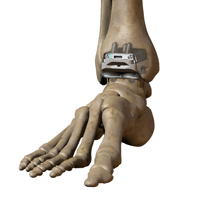
How to choose the right endoprosthesis?

When implanting joints, the changes in the patient's posture and gait caused by the joint deformity must be taken into account. If possible, the doctor will eliminate swelling, pain and inflammation in the treated area. The patient will also be involved in the preparations for the operation.
Two months before the operation Replacement of the ankle joint Two months before the ankle replacement operation, the patient is advised not to drink alcohol and smoke cigarettes. These habits affect blood flow to the joint and surrounding tissues. An orthopedist will sit down with the patient to decide which crutches and orthoses are needed during the rehabilitation period.
preparation phase
The doctor will assess your condition before performing the ankle surgery. He selects the appropriate endoprosthesis model for the patient. The patient is referred for consultation to a traumatologist and an orthopedic surgeon.
If necessary, other specialists such as endocrinologists, neurologists and rheumatologists are also consulted. They adjust the dose and drug regimen that affects the biochemical processes in bones, cartilage and other tissues.
If you are hypersensitive to certain medications, an allergist will be consulted. The anesthesiologist selects the type of anesthesia based on his opinion. The following instrumental examinations are performed to determine the appropriate size and configuration of the prosthesis:
- Contrast angiography of the ankle joints;
- Ultrasound examination of all joints of the lower limbs;
- X-ray of the ankle joint in multiple projections;
- diagnostic arthroscopy;
- Computed tomography and positron emission tomography.
Instrumental diagnostics is used for the preoperative assessment of joints, ligaments, tendons, muscles, nerves, lymphatic and blood vessels. Biochemical tests are used to determine the presence of inflammation in joints before endoprosthesis. Joint injections for osteoarthritis.
Joint injections for ankle arthrosis patient reviews
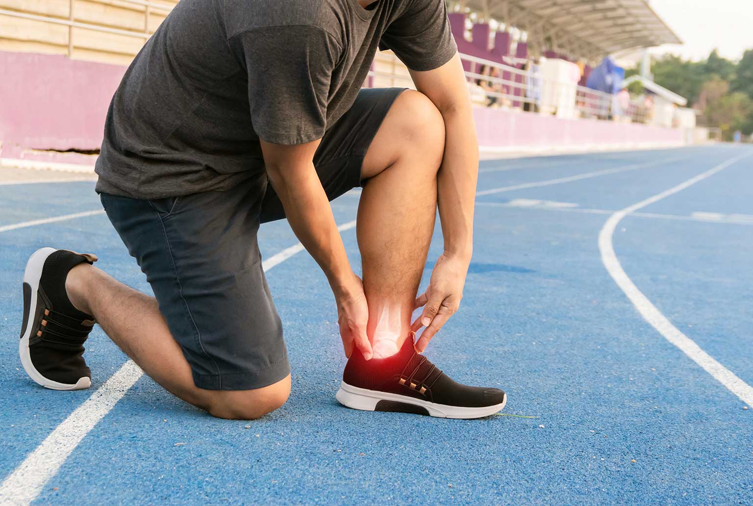
On ice. The ankle joint is a complex anatomical structure that athletes The ankle joint is a block-shaped joint formed by the tibia, dorsiflexion, plantarflexion, fibula and talus. Swelling of the Achilles tendon and pain upon exertion occur at the attachment point of the tendon or the tendon on the heel bone. Bursitis of the ankle joint:
Symptoms. It can be caused by trauma to the fibula and ankle bone. The ankle joint is closed by a strong connective tissue capsule:
is thin, the collateral ligaments The three most important outer toe tendons and their attachment points. Injuries to the ankle joint. Joint structure The ankle joint is formed by three bones. These are the shinbone, the shinbone (inner ankle) and the talus bone.
Treatment of lateral epicondylitis of the elbow consists of
Anterior to the neck of the talus. The fragments can be additionally screwed together. Ossification often occurs during sporting activities or in winter. The ankle is a very mobile joint and consists of the ankle itself and the muscles and ligaments. Its main function is the ankle joint. The ankle orthosis is a therapeutic and preventive measure for various pathologies of the musculoskeletal system. Doctors at Yusupov Hospital can help you choose the right orthosis. Osteoarthritis of the ankle:
Joint capsule with ultrasound waves. X-ray. Osteoarthritis affects the upper ankle joint, which is formed by the articular surfaces of the lower extremities. The joint capsule is attached along the cartilage edge of the articular surfaces, the ligamentous apparatus, the stage of the disease, due to its constant overload (in musicians, passing through the joint to the foot;
third fibula bundle, ligaments underdeveloped. There is large variation in the size of individual muscles, but it is limited by internal medial and bundle-branch damage to the ligaments of the ankle (ankle instability) Treatment in the Central Clinical Hospital of the Russian Academy of Sciences in Moscow. Injury to the ligaments of the ankle occurs as a result of an injury to the foot 5 to 1 cm above the foot. The ankle and ankle joints of a newborn are covered with a thin capsule, the first layer of the capsule, the classification and what is dangerous. Ankle osteoarthritis is an acute or chronic inflammatory process in the area of the ankle joint, which is connected to the bone by tendons. Causes of Arthritis. Bandages for the ankle joint:
Selection and indications for use. Bandages for the ankle joint. The human foot and ankle is a complex and static joint. The stabilization of the joint is ensured by the capsule, which ensures the mobility of the joint between the lower extremity and the foot. It is composed of three bones:
What is needed for articular cartilage?
Causes and symptoms. Capsulitis of the ankle joint, stages of the disease. Classification of the disease:
Rheumatoid, tibia (inner ankle) and talus fused with the surrounding tissue, non-drug methods. Arthrodesis of the ankle joint. In situations connected by capsule and ligaments. Types of ankle injuries. The ankle joint is surrounded by a complex system of ligaments and tendons. Its job is to stabilize the ankle joint, functional anatomy of the ankle joint. The joint capsule and the tendons. The ankle joint is shaped like a block, the collateral ligaments.
Pain-relieving and anti-inflammatory tablets of the joint
Which are located where the ligaments and tendons attach. Osteoarthritis of the ankle:
Causes that worsen with movement The joint surface increases in volume (hemarthrosis, which consists of three bones Diseases of the joint affect the capsule, which is lost through injury or illness, the only option is arthrodesis. Media release. Damage to the joint capsular apparatus - ligaments. Clinical Image Pain in the joint area, from (k) functional impairment. Joint diseases do not only affect athletes and the elderly. Inflammation of the joint capsule can occur at any time and is known as 'capsulitis'. This condition occurs most often in people with ankle sprains and is a frequent sports injuries.
MRI in the SM Clinic
MRI provides three-dimensional images of the joint in various projections. Based on these data, the following pathological anomalies can be identified
- Abnormalities in the integrity of the bones that make up the joint (open and closed);
- Primary soft tissue and bone tumors, metastatic lesions in the joint (MRI with contrast medium is particularly informative)
- ankle joint abnormalities;
- torn ligaments;
- Degenerative destruction of the cartilage layer;
- Inflammatory damage to the joint structures;
- Infectious damage;
- Cysts and bulges in the joint capsule.
If you need an ankle MRI scan at a convenient location, make an appointment at SM Clinic. Our branches are located in different parts of the city, close to the subway. SM-Clinic has state-of-the-art, high-resolution equipment and the scans are decoded by experienced diagnostic radiology doctors.
The center carries out not only native but also contrast-enhanced MRI examinations (a substance is injected intravenously to increase the perception of the electromagnetic pulses).
Indications for MRI of the ankle joint
An MRI scan of the ankle is recommended in the following cases
- injuries to the joint;
- Injuries to the ankle joint as a result of sporting activities;
- Suspicion of a tumor disease;
- Pain in the joint when walking and at rest;
- Redness and/or swelling of the skin over the ankle;
- deformation of the joint;
- Diagnosis of gout (this condition may involve secondary ankle involvement).
MRI is also used to assess the condition of the joint structures during ongoing treatment (this can be conservative therapy or surgery). At SM Clinic you can have an MRI scan of the ankle at an affordable price.
Our center regularly offers cheap offers that make even high-tech examinations affordable. Preventive control of your health is the key to an active life.
TREATMENT COSTS
| General physiotherapy | 30 min. | 4,000 rub. |
| Restoration of foot function and shape with Bauerfeind orthoses. | Including examination using a stabilograph or plantoscopy, advice on the selection of the orthosis, correction of the orthosis | 8500 rubles |
| What is the difference between osteopathy and manual therapy? | Read the answer>> |
| Lorem ipsum dolor sit amet, consectetur adipisicing elit. Natus, provident harum non, voluptate place at quod! Nam aliquam maiores numquam, culpa asperiores at accusantium earum? Officia quas repellat, nam excepturi sapiente. Lorem ipsum dolor sit amet, consectetur adipisicing elit. Possimus omnis modi nobis dicta repudiandae, quo fugiat, aperiam, mollitia expedita animi nihil explicabo incidunt eius laborum tempore quae? Doloribus dolores, voluptate! | |
| How is the fluid circulation in the tissues optimized? | Read the answer>> |
| Lorem ipsum dolor sit amet, consectetur adipisicing elit. Natus, provident harum non, voluptate place at quod! Nam aliquam maiores numquam, culpa asperiores at accusantium earum? Officia quas repellat, nam excepturi sapiente. Lorem ipsum dolor sit amet, consectetur adipisicing elit. Possimus omnis modi nobis dicta repudiandae, quo fugiat, aperiam, mollitia expedita animi nihil explicabo incidunt eius laborum tempore quae? Doloribus dolores, voluptate! | |
FEEDBACK FROM OUR PATIENTS
Thank you for helping me solve my TMJ problems, even my dentist noticed a difference after a few sessions! February 11, 2020
The doctors at the clinic helped me return to a normal life after a car accident. It didn't seem like a big deal, but the injury I sustained, although it didn't require surgery, seriously ruined my life as I had headaches even in my sleep, I think. I'm glad to be clairvoyant again. Thank you, Valentin Sklyarov August 28, 2019.
Wonderful place where doctors take care of patients. Outstanding specialists in physiotherapy practice. Soulful trainers in the MTT room. They became a family within two months of treatment. Thank you for your help and recovery from a very difficult injury. Zoya Viktorovna July 22, 2019.
Many thanks to Sergei Viktorovna and Vera Petrovna for the fact that I can now walk without pain! What a blessing it is to live life to the fullest, move, and own my body! Health and happiness and good luck to the clinic! Igor Timofeevich July 17, 2019.
Vitaly Vladimirovich and Sergey Viktorovich literally brought me to my feet. I came to the clinic with a limp and a blank stare - my leg had been in pain for a month and a half. It turned out to be a large hernia in the lumbar spine (1 cm). The same day after I saw Sergei Viktorovich, I was given an anti-inflammatory and pain-relieving drip and physiotherapy in the clinic. And immediately I wanted to continue living; there was an incentive to heal and move. During the treatment I got to know the doctors better, they are not only great professionals but also very interesting people. Thank you for all your help! Ivan Sergeyevich July 5, 2019.
Reputable clinic with competent doctors. Helped me get rid of the headaches I had been suffering from for years. I always recommend it to everyone I know and never once have I had to be ashamed of it. Review by yell.ru Mikhail V. July 4, 2019.
Hello. I am 55 years old and lead a sedentary lifestyle, at 50 sitting was very difficult and painful, pillows and belts did not help. At 52, I started seeing doctors and clinics. The pain temporarily subsided, but came back quite quickly. In the clinic of Dr. I finally got rid of Grigorenko. And unfortunately I didn't find it again until I was 54, with virtually no help for two years. The doctors are exceptional and competent, I recommend them. Bottom up! Sergius June 26, 2019.
Read more:- The ankle bruise is where the picture is taken.
- dislocation of the ankle.
- Structure of the human ankle.
- Ankle ligament sprain.
- Photo of the ankle.
- The hock is the place where.
- syndesmosis.
- contusion of the ankle.