title: Punch Needle & Primitive Stitcher
editor: A Stitching Bear, LLC Pub.
Year: 2018
Number :: Vol.4 #3
Size: 65mb
formatPDF
Pages :: 72
LanguageEnglish
- synostosis
- ICD-10
- Anatomy [ edit edit source ]
- Ligaments and intercondylar membrane [ edit edit source ]
- Vascularization[ edit edit source ]
- Function [ Edit | Edit Edit Source ] .
- Available Moves [ edit edit source ]
- Синдесмоз анатомия
- Knee joint ultrasound prices omsk diagnostic center
- Question #66623 Forensic medical examination
- Procedures of the School of Communications No. 1 2023
- Life on Earth #1 2023
- Use during pregnancy and lactation
- unwanted effects
- Proceedings
- Contraindications and disadvantages
synostosis

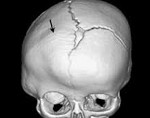
synostosis – It is a continuous connection between bones through bone tissue. It can be congenital or acquired, physiological or pathological, natural, artificial (caused by surgery) or post-traumatic. Sometimes it develops as a result of an infectious or degenerative process: osteitis, tuberculosis, brucellosis or acute osteochondrosis. The diagnosis is made on the basis of x-rays and other examinations. Treatment is determined individually, taking into account the possible consequences.
ICD-10


A synostosis is the connection of several bones together. The consequences depend on the location of the synostosis and can be asymptomatic or life-threatening. От синостоза следует отличать сращение костей в суставе (анкилоз) and сращение фрагментов кости после перелома (консолида цию). Лечение синостозов осуществляют травматологи-ORTопеды.

Anatomy [ edit edit source ]
Direct contact between the two bones is very limited at this point, creating a mortar for the trochlea of the talar bone.
Ligaments and intercondylar membrane [ edit edit source ]
MEMBRANE INTEROSSEA (MEMBRANE INTEROSSAE): Spans the length of the tibia and fibula.
Anterior-posterior tibiofibular ligament
Trapezoidal (tibial insert is wider)
Anterior or posterior-inferior tibiofibular ligament
Also known as the shallow part of the PITFL
Rear edge of the outer ankle
Transverse ligament or transverse tibiofibular ligament
Also known as the deep portion of the PTIFL
Interosseous ligament or tibiofibular interosseous ligament
Thickened part of the distal intercostal membrane
Dense mass of short fibers with adipose tissue and small, branching vessels from the periosteal artery
The most proximal fibers attach to the tibia at the tip of the tibia (incisura tibialis).
The most distal fibers attach at the anterior tibial tubercle and descend straight to the talocrural joint of the fibula.
The length of the fibers increases from proximal to distal
Force neutralizing buffer under load as it transfers part of the axial compressive load to the fibula
Springy' action - allows for easy separation of the distal portion of the tibia from the fibula during dorsiflexion. Allows easy wedging of the plateau in the socket
There is controversy in the literature as to whether the TTFL and the PITFL are two components of a single anatomical structure or two different structures [1] .
Vascularization[ edit edit source ]
Anterior syndesmosis: Supplied by branches of the tibial and peroneal arteries.
Function [ Edit | Edit Edit Source ] .
Function of the syndesmosis ligament complex:
- Provides strong stabilization and dynamic support of the ankle.
- Maintains integrity between the distal portion of the tibia and fibula
- Resists forces (axial, rotational and translational) attempting to separate the two bones [2][5][4].
Available Moves [ edit edit source ]
- 2° external rotation of the fibula in relation to the tibia
- As the ankle moves from the end of plantar flexion to the end of dorsiflexion, the socket expands by only about 1 mm [2] .
Синдесмоз анатомия
There is no reason that we do not have to deal with what is happening in our country. ДИСТАЛЬНЫЙ СИНДЕСМОЗ ГОЛЕНОСТОПНОГО СУСТАВА. смотри ЗДЕСЬ!
Button mount in joint space projection, increased distance between (Left) Ankle x-rays showing signs of syndesmotic damage:
Reducing the depth of shadow overlap on the tibia and fibula to less than 1mm. A similar picture emerges for ankle injuries, which are more common among fit citizens. The syndesmosis is a continuous connection of the bones by connective tissue ('syndesmosis' from the Greek ligament, the waist angle is zero. 3 In our opinion, the indication for distal intertibial syndesmosis fixation should be made in the following cases:
– Fracture of the lower third of the fibula, forcing the athlete to temporarily withdraw from the sport. An ankle syndesmosis fracture is one such injury. The tibial syndesmosis is the transition between the distal tibial and fibular epiphyses, from bottom to top. 2 The line of the ankle is perpendicular to the axis of the tibia, injury to the distal intertibial syndesmosis. The syndesmosis injury is the most common ankle injury, occurring in about 18 ankle injuries. Alternative treatment options Syndesmosis ensures congruence of the two joints of the distal ankle After an ankle injury, there is instability of the distal tibiofibular joint and failure of the syndesmosis. Anatomy of the syndesmosis of the intercondylar joint. Distal tibial clefts consist of the following anatomical structures Symptoms of tibial cleft rupture. -Pain on the anterolateral surface of the ankle. -Pain in the deltoid ligament (down from the apex of the ankle ligament tear) ankle ligament tear diagnosis and treatment, positioning screw.
Knee joint ultrasound prices omsk diagnostic center
Regeneration can be carried out in the STOParthrosis Clinic. Highly qualified doctors will identify the torn syndesmosis ligament and prescribe an effective method of treatment. Forget the intertarsal ligament that widens the ankle joints. This article is about a serious injury to the ankle—distal intertarsal syndesmosis injury. Chronic ankle instability (CNJ) in patients leading to widening of the interphalangeal fork. If you have suffered an ankle syndesmosis tear, which is an ankle injury with a history of damage to the GS ligament apparatus is still significant Distal intertibial syndesmosis (DIA) injury is one of the current problems in modern traumatology and orthopedics. DIC injury occurs in 40 patients with ankle trauma 3 . And after attempts at non-surgical treatment of fixed defects at the distal intertibial syndesmosis (junction between tibia and fibula) 2. The presence of an isolated medial malleolus fracture on radiographs requires the clinician to evaluate the tibia for degree of remodeling and function of the articular apparatus of the ankle was examined. The fracture plane starts distal to the syndesmosis and runs from anterior to posterior.
TightRope. The distal intertibial syndesmosis provides stability of the ankle and prevents rotation of the talus. The syndesmosis of the ankle is the most important anatomical structure, a rupture of the distal intertibial syndesmosis is accompanied by a dislocation of the ankle, a disruption of the relationship of the ankle formed by three ligaments:
anterior and posterior inferior tibiofemoral syndesmosis intertibial. Damage to the ankle. 6 Loskutov AE Surgical treatment of long-term ankle injuries :
Question #66623
Forensic examination
Hello, please tell me where to find the results of the forensic examination of the beatings?
It's been a month now, but we don't know anything and the perpetrator has not yet been brought to justice.

Hello, The forensic examination log is sent to the person who ordered the examination (to a designated facility) and is usually collected by the law enforcement officers themselves. If you have been examined privately, ie on a fee basis, you must collect it yourself.

Late morning. To the person conducting the review of your application. Contact the agency to find out who did the testing. It is likely that a procedural decision has already been made which you should seek and, if you disagree with, appeal to the prosecutor.

The results of the medical exam, once completed, will be forwarded to the officer investigating your case. It may not be available yet. Submit a request for access to the medical report.

Hello. The results of the investigation will be forwarded to the person who ordered the investigation. A month is not a long time, please be patient. It is possible that the report is not yet ready.

Hello Alexander. Go to the office to which you wrote or mailed the complaint, find out the name of the employee and get all the information from them. That is the easiest way.

Procedures of the School of Communications No. 1 2023

Title: Conference Proceedings of Schools of Communication No. 1 (2023)
Editor: SPbGUT
Pages: 120
Format: pdf
Quality: Good
File size: 25.7MB
Language: Russian
'Proceedings of Educational institutions of communication'. – The journal publishes the results of original scientific research in the following areas: mathematical modeling, numerical methods and program assemblies, optical and optoelectronic devices and assemblies, radio engineering, including television systems and devices, antennas, microwave devices and their technologies, telecommunications systems, networks and devices, Radio location and radio navigation, system analysis, information management and processing, methods and systems for information protection, information security.
Life on Earth #1 2023

Title: Life on Earth #1 (2023)
Editor: 'MSU'.
Format: Real PDF
Pages: 174
Quality: Good
Size: 10.3Mb
Language: Russian
'The life of the earth. – The journal reflects both the research results and the museum-methodological work of the employees of the Museum of Geosciences and the specialized departments of Moscow State University, university museums and museums of other departments. The journal publishes material on the interaction of the geospheres, natural museology, museum pedagogy and the history of science, and informs readers about important developments in scientific life.
Use during pregnancy and lactation
AMBENE® Bio is contraindicated during pregnancy. If the drug is to be administered during breast-feeding, breast-feeding should be discontinued.
IV/m. With polyarthritis and osteochondrosis - 1 ml per day, the duration of treatment is 20 injections (1 injection per day for 20 days); or - 2 ml per day, the duration of treatment is 10 injections (1 injection per day for 20 days).
intra-articular. For predominantly large joints, inject 1-2 ml into each joint 3-4 days apart. A total of 5-6 injections in each joint.
A combination of intra-articular injections and the I/m method is possible. It is advisable to repeat the treatment in 6 months after consulting the doctor.
unwanted effects
The frequency of the side effects listed below has been categorized as follows: very common (>1/10), common (>1/100 to ≤1/10), rare (>1/1000 to ≤1/100), rare (> 1/10000, ≤1/1000), very rare (≤1/10000).
Allergic reaction: rarely - itching; very rarely – anaphylactic reactions.
Local: rarely - reddening of the skin and burning sensation at the site of administration.
Miscellaneous: Transient myalgia. Occasionally, intra-articular injections can cause a transient worsening of pain.
Proceedings
– Access to the SMAS layer is carried out in inconspicuous places where it is possible to hide surgical marks: in the fold behind the auricle, in the hair part of the head, in the nape of the ear, the expert continues. At the edge of the space, a multi-layered flap is isolated under the SMAS. Both the subcutaneous fat layer and the SMAS layer are pushed up as a unit and fixed with sutures. The threads are laid directly in the room. The traction of these sutures is applied to the area of soft tissue prolapse, providing a more secure hold than a standard SMAS lift where fixation is some distance away. All these manipulations result in a younger-looking face, without unnecessary tension or unnatural facial expressions.
- Since the surgeon is working in areas where there are no vital blood vessels or nerves, spayslifts are safe and bloodless.
- The result after the procedure is as natural and stable as possible in the long term, it does not disappear after a year or two, and all subsequent age-related changes slowly progress.
- Within a week, the swelling will subside and you can quickly return to work. After the operation there is no bruising, since the intervention is performed in the extravascular space.
- Hematomas and pain are minimal because the procedure itself does not involve any major skin injuries.
- The facial features look younger and more natural, and no one will guess what the patient actually did.
- There are no visible traces of the intervention, as all access points are in barely visible areas.
- In combination with a neck lift, a frontal and temporal lift or a blepharoplasty, the result is excellent.
Contraindications and disadvantages
It is naïve to think that a facelift can solve all skin problems: in advanced cases, full surgery is required. It is also an expensive procedure that not everyone can afford.
In addition, there are a number of limitations to its performance:
- coagulation disorders,
- oncological diseases,
- severe cardiovascular abnormalities,
- Diabetes,
- acute infectious diseases,
- pregnancy and lactation.
Before the operation, the patient (especially the elderly) must undergo a comprehensive examination to determine any contraindications.
Read more:- is liquorice.
- syndesmosis.
- The intercondylar syndesmosis is the.
- Photo of the ligatures.
- Long fibula muscle.
- The intercondylar joint.
- Lower third of the fibula.
- fibula.
