Polymethyl methacrylate (bone cement) is used to cement the prosthesis into the bone. Individual implants may have a specific classification. For example, knee endoprostheses can be unicondylar and are used when only the damaged part of the cartilage can be replaced.
- Upper limb circumference
- Anatomy of the clavicle
- FIXED JOINTS
- FREELY MOVABLE JOINTS
- CONTACT
- Fitness training for trainers.
- Our courses are suitable for both beginners and experienced trainers and offer income growth and career development.
- Types and forms of connections
- Structure of the joint
- Ligaments in the joint
- Structure of the joint in humans
- Joint anatomy: blood supply and innervation
- Functions of the joints
- Classification of the joints
- Specificity of the endoprosthesis of individual joints.
- Replacement of large joints: features of the prosthesis and rehabilitation period
- Where is arthroplasty performed?
- Joint diseases of the hand
- shoulder joint
- elbow joint
- Joint diseases in the legs
- hip joint
- Knee joint
Upper limb circumference
The shoulder girdle is the part of the skeleton that consists of a pair of shoulder blades, the clavicles, and the humerus. The collarbones (clavicles) at the front of the chest and the shoulder blades (scapulae) at the back are connected to the shoulders and support them. The muscles and ligaments ensure the stability and mobility of the skeleton of the upper limbs. The most common pathologies of the shoulder girdle include dislocations, fractures and inflammatory diseases.
The large, flat, triangular bone located on the upper back is called the scapula. It is a paired bone with the base pointing upward and the pointed end pointing downward on either side of the spine. It looks like a wide, flat structure that is slightly curved backwards.
A notice. A pair of shoulder blades at the back and a pair of clavicles at the front are required to form the bony shoulder rim.
- The ostium is the bony ridge that crosses the upper edge of the scapula.
- The acromion is the humerus bone where the neck ends. The connection between the acromion and the clavicle is the acromioclavicular joint.
- The clavicle is a hook-shaped projection in the shape of a bird's beak that sits on top of the bony structure next to the socket. Muscles and ligaments are attached to it.
- The neck is the constricted area adjacent to the socket of the bone. This area serves as a connection between the articular surfaces of the scapula and the shoulder and forms the scapula joint.
- The body of the scapula.
- Side edge.
- External angle.
The anterior surface of the human scapula consists of these elements:
The anterior surface of the bony structure is concave and the posterior surface is convex. The scapular muscle attaches to the front.
The upper edge of the bone contains a cavity in which nerve fibers and blood vessels pass. The vertebral border lies next to the spine. And the lateral edge is the largest area formed by the nodules of the shoulder muscle.
- Upper – has a rounded shape and is located at the top.
- Lower – has a thicker structure.
- Lateral – is strongly thickened and surrounds the joint socket, which is connected to the head of the humerus. It is located opposite the medial superior angle.
Anatomy of the clavicle
The S-shaped bone that curves along its long axis is called the clavicle. It is located horizontally on the front and upper side of the chest. This bone borders the neck. The clavicle is a long bone and consists mainly of spongy material.

The inner end is adjacent to the handle of the sternum and has a bulge that curves forward while the other part is curved backwards. The middle part of the bony structure is slightly compressed from top to bottom. There is a nutrient hole in the lower part of the clavicle. At the inner end there is the recess for the rib-clavicular ligament.
The left outer end of the bone is connected to the acromion of the scapula. This part of the bone has a cone-shaped bulb and also a trapezoidal line. On the underside of the clavicle shaft, closer to the acromion, there is a depression for the insertion of the muscle of the same name.
The bone is smooth on the top and rough on the bottom, with bumps and lines. Its inner end is thicker. The articular surface is located on the inside. The outer end is wider but not as thick. It often connects the clavicle to the acromion of the scapula.
FIXED JOINTS
The joints of the individual bones of the skull and pelvis are examples of rigid, immovable, or otherwise fixed joints. These joints are only mobile during early childhood, during growth or in special cases such as pregnancy. These joints are special because they are not affected by RA.
Some joints, such as B. those between the vertebrae of the spine are only slightly mobile. The bony structures of the vertebrae (spine and cervical spine) are separated by a cartilage pad, the intervertebral disc. When these joints move more than absolutely necessary, problems can occur. An example of this is the well-known 'herniated disc', which can occur when a slightly mobile disc moves further than it should. These joints are also not affected by rheumatoid arthritis.
FREELY MOVABLE JOINTS
Freely movable joints, so-called synovial joints or dart roses. These are the types of joints most people think of when asked to name them. The shoulders, elbows, wrists, fingers and toes, hips, knees and ankles are freely movable joints. The synovial joints can be affected by RA.
There are major differences in structure and function between the various synovial joints. To allow for a wide range of movement, we have ball joints. Knee and elbow joints, for example, can move primarily in one direction because the contours of the bones at the joints fit together like a rod. In contrast, the hip and shoulder joints can move in multiple axes and directions. Although the function of the individual synovial joints is organized differently, they all consist of the same parts.
CONTACT
For comprehensive information on the treatment and prevention of orthopedic, rheumatological or neurological diseases, please contact us:
 Tel. +7(495)120-46-92
 Email: [email protected]
feedback form  Send us a message on Telegram
  Contact us on WhatsApp

Our address is 11 Trifonovskaya St., Moscow
Fitness training for trainers.
For fitness trainers looking for courses that will increase their income, make them even cooler and allow them to never struggle for a job again, we recommend looking into the following distance learning courses:
Basic personal trainer course – For trainers who want to supplement their knowledge with basic information. An incredible amount of useful material that will take you to the next level.
upgrade – An online course for fitness trainers who want to expand their income and knowledge. Tailor-made themes.
architecture of the body – Dmytro Gorkovsky's signature course with full-day practical and theoretical training for fitness trainers, massage therapists and doctors.
pregnancy – Training techniques for pregnancy and recovery courses after birth.
Pilates - Ekaterina Vasilenko's online course on the techniques of the Pilates system.
Our courses are suitable for
Suitable for both beginners and experienced trainers, offering income growth and career development.
The user who purchases services on the website evotren.ru, hereinafter referred to as the 'Customer', on the one hand, and Evotren LLC, hereinafter referred to as the 'Contractor', represented by the Managing Director FG Kapishev, acting within the framework of the Charter, on the other hand, are automatically entered into this contract (hereinafter referred to as the 'Contract') if you purchase the services of the Contractor as follows:
1. TERMS AND DEFINITIONS USED IN THE CONTRACT
1.1. customer – Natural person, private entrepreneur or legal entity, regardless of its organizational and legal form, who places an order with the Contractor in accordance with the terms of this Agreement by purchasing the Contractor's services.
1.2. contractor – The legal entity that provides services to the customer under the contract.
1.3. Services – Services providing access to the study of the distance learning materials listed in the descriptions of the information courses.
1.4. site – The contractor's information resource, located on the Internet at: edu.evotren.com
1.5. Personal customer account – The program interface on the website for studying information materials and other necessary information, to which the customer has access after authorization through a login and password.
1.6. Order – An automatically generated document detailing the services requested by the customer. The order is drawn up by filling out the necessary forms on the website of the realizer -www.evotren.ru.
1.7. Acceptance of Terms and Conditions – the acceptance of the contractual conditions by the customer by paying for the services in cash, non-cash or electronically. Acceptance of the terms and conditions of the contract is deemed to have occurred when the customer pays for the services in cash or non-cash or electronically.
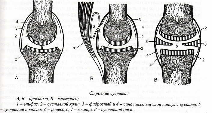
Types and forms of connections
There are simple joints, which consist of two bones, and complex joints, which consist of several elements.
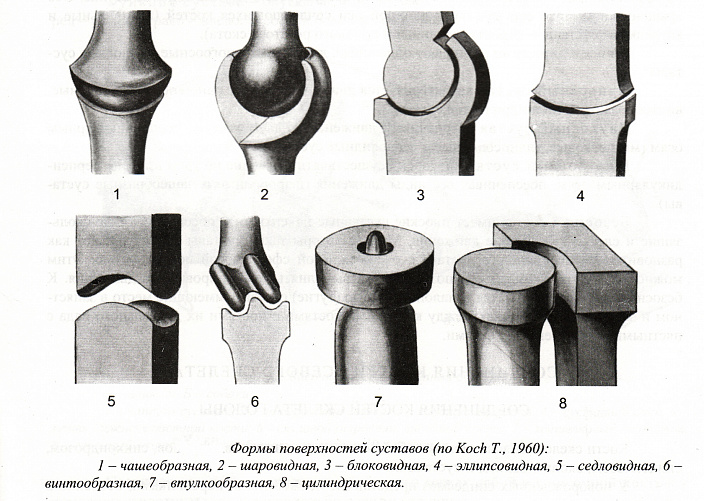
The joints are shaped like geometric figures and therefore have similar names: spherical, ellipsoidal, cylindrical (but there are also block-shaped and saddle-shaped joints).
The shape of the joint determines whether the joint moves around one axis, two axes, or even three axes.
Therefore, there are uniaxial (block and cylindrical), biaxial (saddle and elliptical) and multiaxial (spherical) joints.
The number of axes in a joint depends on the type of movements it is intended to perform. These can include flexion and extension (movement about the front axis), adduction and extension (sagittal axis) and rotational movements (longitudinal axis).
Structure of the joint
A joint is defined as a movable connection between two or more bones. Their anatomical structure is unique and diverse, but we can distinguish them:
- articular surfaces lined with cartilage;
- joint cavities;
- the joint capsule
- the synovial membrane and the fluid within it.
The smoothness of the articular surfaces of the bones that make up a joint is ensured by their constant sliding movement. The cartilage is not only a protective layer, but it also has a shock-absorbing function. The main function of the joint capsule is to protect the joint from external influences. The nerve endings located in the joint capsule also have a good protective function, because pain is the signal that a part of the body or an area sends to the brain to 'decide' what to do. There are menisci in the joint cavity, which protect the joint while allowing it to move.
Ligaments in the joint
The ligaments play an important role in the anatomy of the joint. They are strong connective tissue fibers that evenly support the anatomical structures. They also limit the movements of the bones that make up the joint. To put it simply, the ligaments ensure that our joints do not rotate 360 degrees. However, some large joints require additional support. The hip joint, for example, has an inner layer of ligaments. This is why ligament injuries are so dangerous and can have serious and very serious consequences. In most cases, surgery is required for effective and good treatment.
In order for the joint to function properly, it must be adequately supplied with nutrients, and this can only happen through the blood supply. The joint capsule is surrounded by 3-8 arteries. These transport nutrients and oxygen. The dense network of the venous system ensures the outflow of blood containing metabolic products. To maintain the normal functioning of the joint, its full innervation is required, which is ensured by the intertwining of sympathetic and spinal nerves. Each anatomical area of the joint is innervated, since the activation of the defense system depends on it (remember that pain is a defense mechanism of the body).
Structure of the joint in humans
A joint is a place where two or more bones come together to form a functional system that allows a person to maintain a stable posture and move in space. The main parts of a joint are represented by the following components:
The articular surfaces are located on the connecting bones and are covered by a thin, 0.2 to 0.5 mm thick cartilage. This cartilage has a dense, elastic structure consisting of intertwined hyaline fibers. The completely smooth surface, polished by the constant sliding of the bones against each other, greatly facilitates movement within the joint, and the elastic cartilage provides safety by acting as a kind of shock absorber during stress and sudden impacts.
The joint capsule forms a tight cavity around the joint that protects it from external influences. It consists of elastic threads that are tightly woven together and anchored to the base of the bones that form the joint. Fibers from the adjacent muscles and tendons are woven into the walls of the capsule, which give it its special strength.
The outer part of the joint capsule is surrounded by a fibrous membrane and the inner part by a synovial membrane. The outer fibrous layer is denser and thicker because it consists of elongated bands of fibrous connective tissue. The synovial membrane is less massive. Most of the nerve endings that are responsible for the pain sensitivity of the joint are concentrated here.
The synovial membrane and the articular surfaces form a narrow, gap-shaped space - the joint cavity. It may contain menisci and intervertebral discs that provide mobility and support to the joint.
On the surface of the synovial membrane there are special secretory villi that are responsible for the production of synovial fluid. This substance, which fills the interior of the cavity, nourishes and moisturizes the joint, softening the friction that occurs between the articular surfaces during movements.
Joint anatomy: blood supply and innervation
In order to maintain the physiological performance of a joint, it must be adequately supplied with nutrients, which largely occurs via the circulatory system. The arterial networks surrounding the joint capsule typically consist of three to eight arteries of varying diameters and transport oxygen molecules and nutrients to the tissues. The venous system is responsible for the proper removal of toxins and contaminants from surrounding tissues.
The joint is innervated via the sympathetic nervous system and the spinal nerve plexus. Nerve endings are found in almost all anatomical units that make up the joint, with the exception of hyaline cartilage. Their sensitivity influences the perception of pain and the activation of the body's defense mechanisms.
Functions of the joints
The main function of joints is to connect bone formations into a single structure. Together with the bones and ligaments, they form the passive part of the musculoskeletal system, which moves with the help of muscle fibers. Thanks to the joints, the bones can change their position in relation to each other by sliding rather than rubbing against each other. The slightest thinning of the joint tissue can have serious consequences, as bone structures wear out very quickly due to friction, causing severe pain and irreversible skeletal deformation.
Joints also help to fix the body in space. Immovable joints serve to keep the skull at rest, sedentary joints serve to maintain an upright posture, and movable joints are classified as locomotor organs.
Classification of the joints
In general anatomy, joints are divided into several groups depending on the number and shape of the articular surfaces, the functions performed and the range of motion. Depending on the number of articular surfaces, the following types of joints are distinguished:
Specificity of the endoprosthesis of individual joints.
The prostheses are manufactured taking into account the unique anatomy and biomechanics of each joint. The table shows some of the features of arthroplasty in different areas.
Replacement of large joints: features of the prosthesis and rehabilitation period
Follow-up rehabilitation (examination by a doctor)

If a custom-made prosthesis is required, it will also need to be replaced separately. This is usually accompanied by significant bone loss - these can be congenital or traumatic lesions, consequences of oncological processes and revision procedures due to periprosthetic infection.[6] 3D modeling techniques are used to create these implant components.[7]
Where is arthroplasty performed?
Major joint arthroplasty is widespread in industrialized countries. Current approaches suggest that surgery should be considered for grade 2 osteoarthritis.[8] In Russia, hip and knee replacements are performed in most major cities. Endoprostheses for other areas are also available, but only in specialized medical centers.
The cost of an endoprosthesis and the cost of surgery to replace different joints are very different. For example, in the N. Priorov Institute of Traumatology and Orthopedics, replacement of the toe joint costs RUB 77,940, while replacement of the main joint costs RUB 105,180. This price does not include the cost of the endoprosthesis.

Joint diseases of the hand
shoulder joint
The shoulder joint is the most mobile and loose joint in the human body. It is formed by the head of the humerus and the socket of the scapula. It is strengthened only by the muscles of the upper limbs and therefore is often affected by various damages.
Causes that can cause pain:
- Injuries (muscle or tendon tears)
- Osteoarthritis (wear and tear of joint cartilage and other joint components)
- Osteochondrosis of the cervical spine
- Tendonitis (tendonitis)
- Inflammation of the shoulder nerve
- arthritis (inflammation)
- infections
elbow joint
The elbow joint is formed by the fusion of three bones - the humerus, the radius and the elbow. The weak points of the elbow are the epicondyle of the humerus and the ulnar process. Since the nerve fibers are located near the joint, any injury is accompanied by a strong feeling of pain.
Causes of pain:
- osteochondrosis
- Inflammatory diseases – rheumatoid arthritis, tendinitis, osteoarthritis
- Inflammation of the external epicondyle ('tennis elbow')
- Elbow bursitis (periarticular capsulitis)
- Injuries (dislocations, broken bones)
Joint diseases in the legs
hip joint
The hip joint is the largest joint that connects the pelvis to the lower limbs, and it is also the joint that bears the most weight. This joint is the classic 'hinge' and is formed by the ball head of the femur and the hip socket of the pelvic bone. In addition, part of the femoral neck is part of the socket of the hip joint. This complex arrangement allows freedom and ease of movement.
Causes that can lead to pain:
- Fracture of the femur
- Aseptic necrosis of the femoral head (destruction of the articular part of the bone)
- Arthritis (inflammation of the joints)
- Osteoarthritis (tissue degeneration)
- Bursitis of the spine
- Inflammation of the tendon (tendinitis)
- infections
- injuries
- Tumors
- Damage to the joint caused by rheumatic diseases
Like any other moving joint in the body, the hip joint tends to wear out with age - the cartilage tissue gradually wears away and the articular surfaces of the bones deteriorate. All of this leads to unphysiological friction, which leads to inflammation and pain.
It is also possible that the pain is just a radiation (spread) of the pain in the lower back. Conversely, even if there is an obvious hip injury, the pain may only be felt in the thigh and lower back.
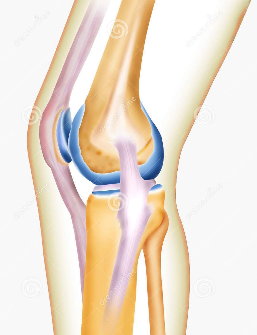
Knee joint
The knee joint is the second largest joint in the body and connects the thigh bone to the lower leg via a series of tendons, muscles and ligaments that stabilize and strengthen the joint.
Knee pain usually indicates damage to one of the components of the joint.
Read more:- Diagram of a joint with and without a dislocation.
- Pronation and supination of the shoulder.
- What is joint rotation?.
- tearing of the joint capsule.
- The pronator muscle - what it means.
- Pronation of the shoulders - what is it?.
- How much does shoulder dislocation surgery cost?.
- Schopar'sche joint.